LI-COR IRDye® 680LT 驴抗鸡二抗,LI-COR IRDye 680LT 二抗

Reagents > IRDye® 680LT Donkey anti-Chicken Secondary Antibody,IRDye® 680LT 驴抗鸡二抗

IRDye® 680LT Donkey anti-Chicken Secondary Antibody

Immunogen

Chicken IgY, whole molecule. IgY is the original designation for the IgG-like protein found in both serum and egg yolk.

Purity and Specificity

The antibody was isolated from antisera by immunoaffinity chromatography using antigens coupled to agarose beads. Based on immunoelectrophoresis and/or ELISA, the antibody reacts with whole molecule chicken IgY, and with the light chains of other chicken immunoglobulins. No reactivity was detected against non-immunoglobulin serum proteins. This antibody was tested by ELISA and/or solid-phase adsorbed to ensure minimal cross-reactivity with bovine, goat, guinea pig, Syrian hamster, horse, human, mouse, rabbit, rat, and sheep serum proteins, but may cross-react with immunoglobulins from other species. The conjugate has been specifically tested and qualified for Western blot applications.

使用与琼脂糖珠偶联的抗原,通过免疫亲和层析从抗血清中分离抗体。 基于免疫电泳和/或 ELISA,抗体与全分子鸡 IgY 以及其他鸡免疫球蛋白的轻链反应。 未检测到针对非免疫球蛋白血清蛋白的反应性。 该抗体通过 ELISA 和/或固相吸附测试,以确保与牛、山羊、豚鼠、叙利亚仓鼠、马、人、小鼠、兔、大鼠和绵羊血清蛋白的交叉反应最小,但可能发生交叉反应 与来自其他物种的免疫球蛋白。 该偶联物经过专门测试,可用于蛋白质印迹应用。

Applications

Recommended for:

  • Western Blot
  • Protein Array
  • Immunohistochemistry
  • Microscopy
  • 2D Gel Detection
  • Tissue Section Imaging
  • 蛋白质印迹
    蛋白质阵列
    免疫组织化学
    显微镜
    2D 凝胶检测
    组织切片成像

Not Recommended for:

  • Small Animal Imaging
  • In-Cell Western Assay
  • On-Cell Western Assay

Formulation

IRDye 680LT secondary antibodies are supplied as purified immunoglobulin conjugates, lyophilized in phosphate-buffered saline, pH 7.4. Protect from light. Store at 4 °C prior to reconstitution.

Each vial contains 10 mg/mL BSA (free of IgG and protease) as a stabilizer and 0.01% sodium azide as a preservative, after reconstitution. Concentration is 1.0 mg/mL when reconstituted as directed. Refer to the pack insert for details on reconstitution.

IRDye 680LT 二抗以纯化的免疫球蛋白偶联物形式提供,在磷酸盐缓冲盐水中冻干,pH 7.4。 避光。 在重组前储存在 4°C。

复溶后,每瓶含有 10 mg/mL BSA(不含 IgG 和蛋白酶)作为稳定剂和 0.01% 叠氮化钠作为防腐剂。 按照指示重新配制时,浓度为 1.0 mg/mL。 有关重组的详细信息,请参阅包装插页。

Recommended Dilutions

Application Recommended Suggested Range
Odyssey Western blot detection 1:20,000 1:20,000 – 1:50,000
Other User optimized

Optimum dilutions will vary and should be determined empirically.

Note: When using PVDF membranes for Western blotting applications, SDS (final concentration of 0.01 – 0.02%) and Tween® 20 (final concentration of 0.1 – 0.2%) must be added during the detection incubation step to avoid non-specific background staining.

RRID

  • P/N 925-68028: RRID AB_2814923
  • P/N 926-68028: RRID AB_10707008

Example Data

IRDye® 680LT Donkey anti-Chicken Secondary Antibody
Western blot analysis of PTEN expression in mouse PTEN transfected 293T whole cell lysate (lane 2) and non-transfected 293T lysate (lane 3). Both lysates were loaded with 2 µg total protein per lane. Odyssey® One-Color Molecular Weight Marker (P/N 928-40000) was loaded in lane 1. The blot was probed with mouse ant-PTEN (IgG2b) (Santa Cruz P/N sc-133197) and chicken anti-GAPDH (ProSci P/N XW-7214) followed by detection with IRDye 800CW Donkey anti-Mouse (P/N 926-32212) and IRDye 680LT Donkey anti-Chicken (P/N 926-68028).
小鼠 PTEN 转染的 293T 全细胞裂解物(泳道 2)和未转染的 293T 裂解物(泳道 3)中 PTEN 表达的蛋白质印迹分析。 两种裂解物每条泳道均加载 2 µg 总蛋白。 将 Odyssey® One-Color Molecular Weight Marker (P/N 928-40000) 加载到泳道 1。用小鼠 ant-PTEN (IgG2b) (Santa Cruz P/N sc-133197) 和鸡抗 GAPDH ( ProSci P/N XW-7214),然后使用 IRDye 800CW 驴防鼠 (P/N 926-32212) 和 IRDye 680LT 驴防鸡 (P/N 926-68028) 进行检测。

LI-COR IRDye® 680LT 驴抗山羊 IgG 二抗,LI-COR IRDye 680LT 二抗

Reagents > IRDye® 680LT Donkey anti-Goat IgG Secondary Antibody,IRDye® 680LT 驴抗山羊 IgG 二抗

IRDye® 680LT Donkey anti-Goat IgG Secondary Antibody

Immunogen

Goat IgG

Purity and Specificity

Isolation of specific antibodies was accomplished by affinity chromatography using goat IgG covalently linked to agarose. Based on ELISA, this antibody reacts with the heavy and light chains of goat IgG and sheep IgG. This antibody has been tested by dot blot and/or solid-phase adsorbed to ensure minimal cross-reactivity with human, mouse, rabbit, rat, chicken. guinea pig, hamster, swine, and horse serum proteins, but may cross-react with immunoglobulins from other species. The conjugate has been specifically tested and qualified for Western blot applications.

使用与琼脂糖共价连接的山羊 IgG 通过亲和层析实现特异性抗体的分离。 基于 ELISA,该抗体与山羊 IgG 和绵羊 IgG 的重链和轻链反应。 该抗体已通过斑点印迹和/或固相吸附测试,以确保与人、小鼠、兔、大鼠、鸡的交叉反应最小。 豚鼠、仓鼠、猪和马血清蛋白,但可能与其他物种的免疫球蛋白发生交叉反应。 该偶联物经过专门测试,可用于蛋白质印迹应用。

Applications

Recommended for:

  • Western Blot
  • Protein Array
  • Immunohistochemistry
  • Microscopy
  • 2D Gel Detection
  • Tissue Section Imaging

Not Recommended for:

  • Small Animal Imaging
  • In-Cell Western Assay
  • On-Cell Western Assay

Note: Do not use this secondary antibody in combination with any secondary antibody whose host is goat (for example, IRDye 800CW Goat anti-Rabbit) for two-color Western blot detection.

Formulation

IRDye 680LT secondary antibodies are supplied as purified immunoglobulin conjugates, lyophilized in phosphate-buffered saline, pH 7.4. Protect from light. Store at 4 °C prior to reconstitution.

Each vial contains 10 mg/mL BSA (free of IgG and protease) as a stabilizer and 0.01% sodium azide as a preservative, after reconstitution. Concentration is 1.0 mg/mL when reconstituted as directed. Refer to the pack insert for details on reconstitution.

IRDye 680LT 二抗以纯化的免疫球蛋白偶联物形式提供,在磷酸盐缓冲盐水中冻干,pH 7.4。 避光。 在重组前储存在 4°C。

复溶后,每瓶含有 10 mg/mL BSA(不含 IgG 和蛋白酶)作为稳定剂和 0.01% 叠氮化钠作为防腐剂。 按照指示重新配制时,浓度为 1.0 mg/mL。 有关重组的详细信息,请参阅包装插页。

Recommended Dilutions

Application Recommended Suggested Range
Odyssey Western blot detection 1:20,000 1:20,000 – 1:50,000
Other User optimized

Optimum dilutions will vary and should be determined empirically.

Note: When using PVDF membranes for Western blotting applications, SDS (final concentration of 0.01 – 0.02%) and Tween® 20 (final concentration of 0.1 – 0.2%) must be added during the detection incubation step to avoid non-specific background staining.

RRID

  • P/N 925-68024: RRID AB_2814908
  • P/N 926-68024: RRID AB_10706168

Example Data

IRDye® 680LT Donkey anti-Goat IgG Secondary Antibody
Western blot detection of actin in C32 lysate. C32 lysate (Santa Cruz P/N sc- 2205) was resolved on a 10% Bis-Tris gel and transferred to Odyssey® nitrocellulose (P/N 926-31090). The membrane was probed with goat anti-actin (Santa Cruz P/N sc-1616) followed by detection with IRDye 680LT Donkey anti-Goat IgG (P/N 926-68024).
蛋白质印迹检测 C32 裂解物中的肌动蛋白。 C32 裂解物 (Santa Cruz P/N sc-2205) 在 10% Bis-Tris 凝胶上分离并转移到 Odyssey® 硝酸纤维素 (P/N 926-31090)。 用山羊抗肌动蛋白 (Santa Cruz P/N sc-1616) 探测膜,然后用 IRDye 680LT 驴抗山羊 IgG (P/N 926-68024) 检测。

LI-COR IRDye® 680LT 驴抗小鼠 IgG 二抗,LI-COR IRDye 680LT 二抗

Reagents > IRDye® 680LT Donkey anti-Mouse IgG Secondary Antibody,IRDye® 680LT 驴抗小鼠 IgG 二抗

IRDye® 680LT Donkey anti-Mouse IgG Secondary Antibody

Immunogen

Mouse IgG

Purity and Specificity

The antibody was isolated by affinity chromatography using antigens coupled to agarose beads. Based on ELISA, this antibody reacts with the heavy and light chains of mouse IgG, and with the light chains of mouse IgM and IgA. This antibody was tested by ELISA and/or solid-phase adsorbed to ensure minimal cross-reactivity with bovine, chicken, goat, guinea pig, horse, human, rabbit, and sheep serum proteins, but may cross-react with immunoglobulins from other species. The conjugate has been specifically tested and qualified for Western blot applications.

使用与琼脂糖珠偶联的抗原通过亲和层析分离抗体。 基于 ELISA,该抗体与小鼠 IgG 的重链和轻链以及小鼠 IgM 和 IgA 的轻链反应。 该抗体通过 ELISA 和/或固相吸附测试,以确保与牛、鸡、山羊、豚鼠、马、人、兔和绵羊血清蛋白的交叉反应最小,但可能与来自其他物种的免疫球蛋白交叉反应 . 该偶联物经过专门测试,可用于蛋白质印迹应用。

Applications

Recommended for:

  • Western Blot
  • Protein Array
  • Immunohistochemistry
  • Microscopy
  • 2D Gel Detection
  • Tissue Section Imaging
  • 蛋白质印迹
    蛋白质阵列
    免疫组织化学
    显微镜
    2D 凝胶检测
    组织切片成像

Not Recommended for:

  • Small Animal Imaging
  • In-Cell Western Assay
  • On-Cell Western Assay
Note: IRDye 680LT secondary antibodies are not intended for In-Cell Western™ assays or in vivo imaging applications.

Formulation

IRDye 680LT secondary antibodies are supplied as purified immunoglobulin conjugates, lyophilized in phosphate-buffered saline, pH 7.4. Protect from light. Store at 4 °C prior to reconstitution.

Each vial contains 10 mg/mL BSA (free of IgG and protease) as a stabilizer and 0.01% sodium azide as a preservative, after reconstitution. Concentration is 1.0 mg/mL when reconstituted as directed. Refer to the pack insert for details on reconstitution.

IRDye 680LT 二抗以纯化的免疫球蛋白偶联物形式提供,在磷酸盐缓冲盐水中冻干,pH 7.4。 避光。 在重组前储存在 4°C。

复溶后,每瓶含有 10 mg/mL BSA(不含 IgG 和蛋白酶)作为稳定剂和 0.01% 叠氮化钠作为防腐剂。 按照指示重新配制时,浓度为 1.0 mg/mL。 有关重组的详细信息,请参阅包装插页。

Recommended Dilutions

Application Recommended Suggested Range
Odyssey Western blot detection 1:20,000 1:20,000 – 1:50,000
Other User optimized

Optimum dilutions will vary and should be determined empirically.

Note: When using PVDF membranes for Western blotting applications, SDS (final concentration of 0.01 – 0.02%) and Tween® 20 (final concentration of 0.1 – 0.2%) must be added during the detection incubation step to avoid non-specific background staining.

RRID

  • P/N 925-68022: RRID AB_2814906
  • P/N 926-68022: RRID AB_10715072

Example Data

IRDye® 680LT Donkey anti-Mouse IgG Secondary Antibody
Western blot detection of actin in C32 lysate. Samples loaded in 4X Protein Sample Loading Buffer (P/N 928-40004), resolved on a 10% Bis-Tris gel, and transferred to Odyssey® nitrocellulose (P/N 926-31090). The membrane was probed with mouse anti-actin (Neomarkers P/N MS-1295) followed by detection with IRDye 680LT Donkey anti-Mouse IgG (P/N 926-68022).

LI-COR IRDye® 680LT 驴抗兔 IgG 二抗,LI-COR IRDye 680LT 二抗

Reagents > IRDye® 680LT Donkey anti-Rabbit IgG Secondary Antibody,IRDye® 680LT 驴抗兔 IgG 二抗

IRDye® 680LT Donkey anti-Rabbit IgG Secondary Antibody

Immunogen

Rabbit IgG, whole molecule

Purity and Specificity

The antibody was isolated from antisera by immunoaffinity chromatography using antigens coupled to agarose beads. Based on immunoelectrophoresis, this antibody reacts with the heavy chains of rabbit IgG, and with the light chains common to most rabbit immunoglobulins. No reactivity was detected against non-immunoglobulin serum proteins. This antibody was tested by ELISA and/or solid-phase adsorbed to ensure minimal cross-reactivity with bovine, chicken, goat, guinea pig, Syrian hamster, horse, human, mouse, rat, and sheep serum proteins, but may cross-react with immunoglobulins from other species. The conjugate has been specifically tested and qualified for Western blot applications.

使用与琼脂糖珠偶联的抗原,通过免疫亲和层析从抗血清中分离抗体。 基于免疫电泳,该抗体与兔 IgG 的重链以及大多数兔免疫球蛋白常见的轻链反应。 未检测到针对非免疫球蛋白血清蛋白的反应性。 该抗体经过 ELISA 和/或固相吸附测试,以确保与牛、鸡、山羊、豚鼠、叙利亚仓鼠、马、人、小鼠、大鼠和绵羊血清蛋白的交叉反应最小,但可能发生交叉反应 与来自其他物种的免疫球蛋白。 该偶联物经过专门测试,可用于蛋白质印迹应用。

Applications

Recommended for:

  • Western Blot
  • Protein Array
  • Immunohistochemistry
  • Microscopy
  • 2D Gel Detection
  • Tissue Section Imaging

Not Recommended for:

  • Small Animal Imaging
  • In-Cell Western Assay
  • On-Cell Western Assay

Formulation

IRDye 680LT secondary antibodies are supplied as purified immunoglobulin conjugates, lyophilized in phosphate-buffered saline, pH 7.4. Protect from light. Store at 4 °C prior to reconstitution.

Each vial contains 10 mg/mL BSA (free of IgG and protease) as a stabilizer and 0.01% sodium azide as a preservative, after reconstitution. Concentration is 1.0 mg/mL when reconstituted as directed. Refer to the pack insert for details on reconstitution.

IRDye 680LT 二抗以纯化的免疫球蛋白偶联物形式提供,在磷酸盐缓冲盐水中冻干,pH 7.4。 避光。 在重组前储存在 4°C。

复溶后,每瓶含有 10 mg/mL BSA(不含 IgG 和蛋白酶)作为稳定剂和 0.01% 叠氮化钠作为防腐剂。 按照指示重新配制时,浓度为 1.0 mg/mL。 有关重组的详细信息,请参阅包装插页。

Recommended Dilutions

Application Recommended Suggested Range
Odyssey Western blot detection 1:20,000 1:20,000 – 1:50,000
Other User optimized

Optimum dilutions will vary and should be determined empirically.

Note: When using PVDF membranes for Western blotting applications, SDS (final concentration of 0.01 – 0.02%) and Tween® 20 (final concentration of 0.1 – 0.2%) must be added during the detection incubation step to avoid non-specific background staining.

RRID

  • P/N 925-68023: RRID AB_2814907
  • P/N 926-68023: RRID AB_10706167

LI-COR IRDye® 680LT 山羊抗人 IgG 二抗,LI-COR IRDye 680LT 二抗

Reagents > IRDye® 680LT Goat anti-Human IgG Secondary Antibody,IRDye® 680LT 山羊抗人 IgG 二抗

IRDye® 680LT Goat anti-Human IgG Secondary Antibody

Immunogen

Human IgG

Purity and Specificity

Isolation of specific antibodies was accomplished by affinity chromatography using human IgG covalently linked to agarose. Based on ELISA, this antibody reacts with the heavy and light chains of human IgG. This antibody was tested by dot blot and/or solid-phase adsorbed to ensure minimal cross-reactivity with bovine, horse, and mouse serum proteins, but may cross-react with immunoglobulins from other species. The conjugate has been specifically tested and qualified for Western blot applications.

使用与琼脂糖共价连接的人 IgG 通过亲和层析实现特异性抗体的分离。 基于 ELISA,该抗体与人 IgG 的重链和轻链反应。 该抗体通过斑点印迹和/或固相吸附测试,以确保与牛、马和小鼠血清蛋白的交叉反应最小,但可能与来自其他物种的免疫球蛋白交叉反应。 该偶联物经过专门测试,可用于蛋白质印迹应用。

Applications

Recommended for:

  • Western Blot
  • Protein Array
  • Immunohistochemistry
  • Microscopy
  • 2D Gel Detection
  • Tissue Section Imaging
  • 蛋白质印迹
    蛋白质阵列
    免疫组织化学
    显微镜
    2D 凝胶检测
    组织切片成像

Not Recommended for:

  • Small Animal Imaging
  • In-Cell Western Assay
  • On-Cell Western Assay

Formulation

IRDye 680LT secondary antibodies are supplied as purified immunoglobulin conjugates, lyophilized in phosphate-buffered saline, pH 7.4. Protect from light. Store at 4 °C prior to reconstitution.

Each vial contains 10 mg/mL BSA (free of IgG and protease) as a stabilizer and 0.01% sodium azide as a preservative, after reconstitution. Concentration is 1.0 mg/mL when reconstituted as directed. Refer to the pack insert for details on reconstitution.

IRDye 680LT 二抗以纯化的免疫球蛋白偶联物形式提供,在磷酸盐缓冲盐水中冻干,pH 7.4。 避光。 在重组前储存在 4°C。

复溶后,每瓶含有 10 mg/mL BSA(不含 IgG 和蛋白酶)作为稳定剂和 0.01% 叠氮化钠作为防腐剂。 按照指示重新配制时,浓度为 1.0 mg/mL。 有关重组的详细信息,请参阅包装插页。

Recommended Dilutions

Application Recommended Suggested Range
Odyssey Western blot detection 1:20,000 1:20,000 – 1:50,000
Other User optimized

Optimum dilutions will vary and should be determined empirically.

Note: When using PVDF membranes for Western blotting applications, SDS (final concentration of 0.01 – 0.02%) and Tween® 20 (final concentration of 0.1 – 0.2%) must be added during the detection incubation step to avoid non-specific background staining.

RRID

  • P/N 925-68032: RRID AB_2814911
  • P/N 926-68032: RRID AB_10795013

LI-COR IRDye® 680LT 山羊抗小鼠 IgG 二抗,LI-COR IRDye 680LT 二抗

Reagents > IRDye® 680LT Goat anti-Mouse IgG Secondary Antibody,IRDye® 680LT 山羊抗小鼠 IgG 二抗

IRDye® 680LT Goat anti-Mouse IgG Secondary Antibody

Immunogen

Mouse IgG paraproteins

Purity and Specificity

Isolation of specific antibodies was accomplished by affinity chromatography using pooled mouse IgG covalently linked to agarose. Based on ELISA and flow cytometry, this antibody reacts with the heavy and light chains of mouse IgG1, IgG2a, IgG2b, and IgG3, and with the light chains of mouse IgM and IgA. This antibody was tested by dot blot and and/or solid-phase adsorbed for minimal cross-reactivity with human, rabbit, goat, rat, and horse serum proteins, but may cross-react with immunoglobulins from other species. The conjugate has been specifically tested and qualified for Western blot applications.

使用与琼脂糖共价连接的混合小鼠 IgG 通过亲和层析实现特异性抗体的分离。 基于 ELISA 和流式细胞术,该抗体与小鼠 IgG1、IgG2a、IgG2b 和 IgG3 的重链和轻链以及小鼠 IgM 和 IgA 的轻链反应。 通过斑点印迹和/或固相吸附测试该抗体与人、兔、山羊、大鼠和马血清蛋白的交叉反应最小,但可能与来自其他物种的免疫球蛋白交叉反应。 该偶联物经过专门测试,可用于蛋白质印迹应用。

Applications

Recommended for:

  • Western Blot
  • Protein Array
  • Immunohistochemistry
  • Microscopy
  • 2D Gel Detection
  • Tissue Section Imaging
  • 蛋白质印迹
    蛋白质阵列
    免疫组织化学
    显微镜
    2D 凝胶检测
    组织切片成像

Not Recommended for:

  • Small Animal Imaging
  • In-Cell Western Assay
  • On-Cell Western Assay

Formulation

IRDye 680LT secondary antibodies are supplied as purified immunoglobulin conjugates, lyophilized in phosphate-buffered saline, pH 7.4. Protect from light. Store at 4 °C prior to reconstitution.

Each vial contains 10 mg/mL BSA (free of IgG and protease) as a stabilizer and 0.01% sodium azide as a preservative, after reconstitution. Concentration is 1.0 mg/mL when reconstituted as directed. Refer to the pack insert for details on reconstitution.

IRDye 680LT 二抗以纯化的免疫球蛋白偶联物形式提供,在磷酸盐缓冲盐水中冻干,pH 7.4。 避光。 在重组前储存在 4°C。

复溶后,每瓶含有 10 mg/mL BSA(不含 IgG 和蛋白酶)作为稳定剂和 0.01% 叠氮化钠作为防腐剂。 按照指示重新配制时,浓度为 1.0 mg/mL。 有关重组的详细信息,请参阅包装插页。

Recommended Dilutions

Application Recommended Suggested Range
Odyssey Western blot detection 1:20,000 1:20,000 – 1:50,000
Other User optimized

Optimum dilutions will vary and should be determined empirically.

Note: When using PVDF membranes for Western blotting applications, SDS (final concentration of 0.01 – 0.02%) and Tween® 20 (final concentration of 0.1 – 0.2%) must be added during the detection incubation step to avoid non-specific background staining.

RRID

  • P/N 925-68020: RRID AB_2687826
  • P/N 926-68020: RRID AB_10706161

Example Data

IRDye® 680LT Goat anti-Mouse IgG Secondary Antibody
Western blot detection of ERK2 in Hela lysate (5 µg – 78 ng). Samples loaded in 4X protein loading buffer (P/N 928-40004), resolved on a 10% Bis-Tris gel, and transferred to Odyssey® nitrocellulose (P/N 926-31090). The membrane was probed with mouse anti-ERK2 (Santa Cruz P/N sc-1647) followed by detection with IRDye 680LT Goat anti-Mouse IgG (P/N 926-68020).
对 Hela 裂解液 (5 µg – 78 ng) 中的 ERK2 进行蛋白质印迹检测。 样品上样于 4X 蛋白上样缓冲液 (P/N 928-40004),在 10% Bis-Tris 凝胶上分离,然后转移至 Odyssey® 硝酸纤维素 (P/N 926-31090)。 用小鼠抗 ERK2 (Santa Cruz P/N sc-1647) 探测膜,然后用 IRDye 680LT 山羊抗小鼠 IgG (P/N 926-68020) 检测。

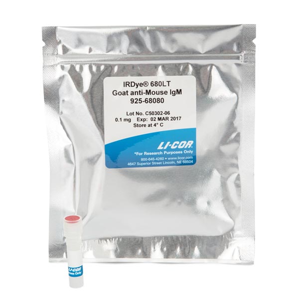
LI-COR IRDye 680LT 二抗,IRDye® 680LT 山羊抗小鼠 IgM(μ 链特异性)二抗

Reagents > IRDye® 680LT Goat anti-Mouse IgM (µ chain specific) Secondary Antibody,IRDye® 680LT 山羊抗小鼠 IgM(μ 链特异性)二抗

IRDye® 680LT Goat anti-Mouse IgM (µ chain specific) Secondary Antibody

Immunogen

Mouse IgM paraproteins

Purity and Specificity

Isolation of specific antibodies was accomplished by affinity chromatography using mouse IgM covalently linked to agarose. Based on ELISA and/or flow cytometry, this antibody reacts with the heavy chain of mouse IgM. This antibody has been tested by dot blot and/or solid-phase adsorbed to ensure minimal cross-reactivity with mouse IgG1, IgG2a, IgG3, and IgA, pooled human sera and purified human paraproteins. The conjugate has been specifically tested and qualified for Western blot applications.

使用与琼脂糖共价连接的小鼠 IgM 通过亲和层析实现特异性抗体的分离。 基于 ELISA 和/或流式细胞术,该抗体与小鼠 IgM 的重链发生反应。 该抗体已通过斑点印迹和/或固相吸附测试,以确保与小鼠 IgG1、IgG2a、IgG3 和 IgA、合并的人血清和纯化的人副蛋白的交叉反应最小。 该偶联物经过专门测试,可用于蛋白质印迹应用。

Applications

Recommended for:

  • Western Blot
  • Protein Array
  • Immunohistochemistry
  • Microscopy
  • 2D Gel Detection
  • Tissue Section Imaging
  • 蛋白质印迹
    蛋白质阵列
    免疫组织化学
    显微镜
    2D 凝胶检测
    组织切片成像

Not Recommended for:

  • Small Animal Imaging
  • In-Cell Western Assay
  • On-Cell Western Assay

Formulation

IRDye 680LT secondary antibodies are supplied as purified immunoglobulin conjugates, lyophilized in phosphate-buffered saline, pH 7.4. Protect from light. Store at 4 °C prior to reconstitution.

Each vial contains 10 mg/mL BSA (free of IgG and protease) as a stabilizer and 0.01% sodium azide as a preservative, after reconstitution. Concentration is 1.0 mg/mL when reconstituted as directed. Refer to the pack insert for details on reconstitution.

IRDye 680LT 二抗以纯化的免疫球蛋白偶联物形式提供,在磷酸盐缓冲盐水中冻干,pH 7.4。 避光。 在重组前储存在 4°C。

复溶后,每瓶含有 10 mg/mL BSA(不含 IgG 和蛋白酶)作为稳定剂和 0.01% 叠氮化钠作为防腐剂。 按照指示重新配制时,浓度为 1.0 mg/mL。 有关重组的详细信息,请参阅包装插页。

Recommended Dilutions

Application Recommended Suggested Range
Odyssey Western blot detection 1:20,000 1:20,000 – 1:50,000
Other User optimized

Optimum dilutions will vary and should be determined empirically.

Note: When using PVDF membranes for Western blotting applications, SDS (final concentration of 0.01 – 0.02%) and Tween® 20 (final concentration of 0.1 – 0.2%) must be added during the detection incubation step to avoid non-specific background staining.
注意:当使用 PVDF 膜进行蛋白质印迹应用时,必须在检测孵育步骤中添加 SDS(终浓度为 0.01 – 0.02%)和 Tween® 20(终浓度为 0.1 – 0.2%)以避免非特异性背景染色。

RRID

  • P/N 925-68080: RRID AB_2814917
  • P/N 926-68080: RRID AB_2814920

Example Data

IRDye® 680LT Goat anti-Mouse IgM (µ chain specific) Secondary Antibody
Western blot detection of guinea pig IgG with IRDye 680LT Donkey anti-Guinea Pig. Serial dilutions of guinea pig IgG were spiked into C32 lysate (Santa Cruz P/N sc-2205). Samples were resolved on a 10% Bis-Tris gel and transferred to Odyssey® Nitrocellulose membrane (P/N 926-31092). The membrane was blocked with Odyssey Blocking Buffer (P/N 927-40000) followed by detection with IRDye 680LT Donkey anti-Guinea Pig (P/N 926-68030).
用 IRDye 680LT Donkey anti-Guinea Pig 对豚鼠 IgG 进行蛋白质印迹检测。 将连续稀释的豚鼠 IgG 加入 C32 裂解液 (Santa Cruz P/N sc-2205)。 样品在 10% Bis-Tris 凝胶上分离并转移到 Odyssey® 硝酸纤维素膜(P/N 926-31092)上。 用 Odyssey 封闭缓冲液 (P/N 927-40000) 封闭膜,然后用 IRDye 680LT 驴抗豚鼠 (P/N 926-68030) 检测。

LI-COR IRDye® 680LT 山羊抗兔 IgG 二抗,LI-COR IRDye 680LT 二抗

Reagents > IRDye® 680LT Goat anti-Rabbit IgG Secondary Antibody,IRDye® 680LT 山羊抗兔 IgG 二抗

IRDye® 680LT Goat anti-Rabbit IgG Secondary Antibody

Immunogen

Rabbit IgG

Purity and Specificity

Isolation of specific antibodies was accomplished by affinity chromatography using pooled rabbit IgG covalently linked to agarose. Based on ELISA and flow cytometry, this antibody reacts with the heavy and light chains of rabbit IgG, and with the light chains of rabbit IgM and IgA. This antibody was tested by dot blot and and/or solid-phase adsorbed for minimal cross-reactivity with human, mouse, rat, sheep, and chicken serum proteins, but may cross-react with immunoglobulins from other species. The conjugate has been specifically tested and qualified for Western blot applications.

使用与琼脂糖共价连接的混合兔 IgG,通过亲和层析实现特异性抗体的分离。 基于 ELISA 和流式细胞术,该抗体与兔 IgG 的重链和轻链以及兔 IgM 和 IgA 的轻链反应。 通过斑点印迹和/或固相吸附测试该抗体与人、小鼠、大鼠、绵羊和鸡血清蛋白的交叉反应最小,但可能与来自其他物种的免疫球蛋白交叉反应。 该偶联物经过专门测试,可用于蛋白质印迹应用。

Applications

Recommended for:

  • Western Blot
  • Protein Array
  • Immunohistochemistry
  • Microscopy
  • 2D Gel Detection
  • Tissue Section Imaging
  • 蛋白质印迹
    蛋白质阵列
    免疫组织化学
    显微镜
    2D 凝胶检测
    组织切片成像

Not Recommended for:

  • Small Animal Imaging
  • In-Cell Western Assay
  • On-Cell Western Assay

Formulation

IRDye 680LT secondary antibodies are supplied as purified immunoglobulin conjugates, lyophilized in phosphate-buffered saline, pH 7.4. Protect from light. Store at 4 °C prior to reconstitution.

Each vial contains 10 mg/mL BSA (free of IgG and protease) as a stabilizer and 0.01% sodium azide as a preservative, after reconstitution. Concentration is 1.0 mg/mL when reconstituted as directed. Refer to the pack insert for details on reconstitution.

IRDye 680LT 二抗以纯化的免疫球蛋白偶联物形式提供,在磷酸盐缓冲盐水中冻干,pH 7.4。 避光。 在重组前储存在 4°C。

复溶后,每瓶含有 10 mg/mL BSA(不含 IgG 和蛋白酶)作为稳定剂和 0.01% 叠氮化钠作为防腐剂。 按照指示重新配制时,浓度为 1.0 mg/mL。 有关重组的详细信息,请参阅包装插页。

Recommended Dilutions

Application Recommended Suggested Range
Odyssey Western blot detection 1:20,000 1:20,000 – 1:50,000
Other User optimized

Optimum dilutions will vary and should be determined empirically.

Note: When using PVDF membranes for Western blotting applications, SDS (final concentration of 0.01 – 0.02%) and Tween® 20 (final concentration of 0.1 – 0.2%) must be added during the detection incubation step to avoid non-specific background staining.
注意:当使用 PVDF 膜进行蛋白质印迹应用时,必须在检测孵育步骤中添加 SDS(终浓度为 0.01 – 0.02%)和 Tween® 20(终浓度为 0.1 – 0.2%)以避免非特异性背景染色。

RRID

  • P/N 925-68021: RRID AB_2713919
  • P/N 926-68021: RRID AB_10706309

Example Data

IRDye® 680LT Goat anti-Rabbit IgG Secondary Antibody
Western blot detection of p38 in Jurkat lysate. Samples loaded in 4X Protein Sample Loading Buffer (P/N 928-40004), resolved on a 10% Bis-Tris gel and transferred to Odyssey® nitrocellulose (P/N 926-31090). The membrane was blocked with Odyssey Blocking Buffer (P/N 927-40000) and probed with rabbit anti-p38 (Santa Cruz P/N sc-7149) followed by detection with IRDye 680LT Goat anti-Rabbit IgG (P/N 926-68021).
Jurkat 裂解物中 p38 的蛋白质印迹检测。 将样品装入 4X 蛋白质样品上样缓冲液 (P/N 928-40004),在 10% Bis-Tris 凝胶上分离,然后转移到 Odyssey® 硝酸纤维素 (P/N 926-31090)。 用 Odyssey 封闭缓冲液 (P/N 927-40000) 封闭膜,并用兔抗 p38 (Santa Cruz P/N sc-7149) 探测,然后用 IRDye 680LT 山羊抗兔 IgG (P/N 926- 68021)。

LI-COR IRDye® 680LT 山羊抗大鼠 IgG 二抗,LI-COR IRDye 680LT 二抗

Reagents > IRDye® 680LT Goat anti-Rat IgG Secondary Antibody,IRDye® 680LT 山羊抗大鼠 IgG 二抗

IRDye® 680LT Goat anti-Rat IgG Secondary Antibody

Immunogen

Rat IgG, whole molecule

Purity and Specificity

The antibody was purified from antisera by immunoaffinity chromatography using antigens coupled to agarose beads. Based on immunoelectrophoresis and/or ELISA, this antibody reacts with whole molecule rat IgG, and with the light chains of other rat immunoglobulins. No reactivity was detected against non-immunoglobulin serum proteins. This antibody has been tested by ELISA and and/or solid-phase adsorbed to ensure minimal cross-reactivity with mouse, bovine, horse, human, and rabbit serum proteins, but may cross-react with immunoglobulins from other species. The conjugate has been specifically tested and qualified for Western blot applications.

使用与琼脂糖珠偶联的抗原,通过免疫亲和层析从抗血清中纯化抗体。 基于免疫电泳和/或 ELISA,该抗体与全分子大鼠 IgG 以及其他大鼠免疫球蛋白的轻链发生反应。 未检测到针对非免疫球蛋白血清蛋白的反应性。 该抗体已通过 ELISA 和/或固相吸附测试,以确保与小鼠、牛、马、人类和兔血清蛋白的交叉反应最小,但可能与其他物种的免疫球蛋白交叉反应。 该偶联物经过专门测试,可用于蛋白质印迹应用。

Applications

Recommended for:

  • Western Blot
  • Protein Array
  • Immunohistochemistry
  • Microscopy
  • 2D Gel Detection
  • Tissue Section Imaging
  • 蛋白质印迹
    蛋白质阵列
    免疫组织化学
    显微镜
    2D 凝胶检测
    组织切片成像

Not Recommended for:

  • Small Animal Imaging
  • In-Cell Western Assay
  • On-Cell Western Assay

Formulation

IRDye 680LT secondary antibodies are supplied as purified immunoglobulin conjugates, lyophilized in phosphate-buffered saline, pH 7.4. Protect from light. Store at 4 °C prior to reconstitution.

Each vial contains 10 mg/mL BSA (free of IgG and protease) as a stabilizer and 0.01% sodium azide as a preservative, after reconstitution. Concentration is 1.0 mg/mL when reconstituted as directed. Refer to the pack insert for details on reconstitution.

IRDye 680LT 二抗以纯化的免疫球蛋白偶联物形式提供,在磷酸盐缓冲盐水中冻干,pH 7.4。 避光。 在重组前储存在 4°C。

复溶后,每瓶含有 10 mg/mL BSA(不含 IgG 和蛋白酶)作为稳定剂和 0.01% 叠氮化钠作为防腐剂。 按照指示重新配制时,浓度为 1.0 mg/mL。 有关重组的详细信息,请参阅包装插页。

Recommended Dilutions

Application Recommended Suggested Range
Odyssey Western blot detection 1:20,000 1:20,000 – 1:50,000
Other User optimized

Optimum dilutions will vary and should be determined empirically.

Note: When using PVDF membranes for Western blotting applications, SDS (final concentration of 0.01 – 0.02%) and Tween® 20 (final concentration of 0.1 – 0.2%) must be added during the detection incubation step to avoid non-specific background staining.

RRID

  • P/N 925-68029: RRID AB_2814909
  • P/N 926-68029: RRID AB_10715073

Example Data

IRDye® 680LT Goat anti-Rat IgG Secondary Antibody
Western blot detection of tubulin in C32 lysate. Samples loaded in 4X Protein Sample Loading Buffer (P/N 928-40004), resolved on a 10% Bis-Tris gel, and transferred to Odyssey® nitrocellulose (P/N 926-31090). The membrane was blocked with Odyssey Blocking Buffer (P/N 927-40000) and probed with rat anti-tubulin (Novus Biologicals P/N NB600-506) followed by detection with IRDye 680LT Goat anti-Rat IgG (P/N 926-68029).