Pearl™ Imager近红外小动物成像系统,926-08446,929-90010,929-80020

系统简介
将成像老鼠放在独特的滑轮式成像床上,轻按成像床关闭按钮。30秒后,无需选择曝光的时间,您就能得到清晰的、三色的小鼠成像。

Pearl™ Imager是美国LI-COR公司专门设计的在近红外波段成像的小动物活体成像系统,专利的基于CCD的光学设计保证红外标记的光学探针具有最优化的成像效果。商品化的光学探针包括IRDye800CW EGF Optical Probe、IRDye800CW 2-DG Optical Probe 、IRDye680/800CW BoneTag和IRDye800CW RGD Optical Probe等。Pearl™ Imager成像系统独一无二的光学设计和优化的光学试剂降低了组织的自发荧光,提供了信噪比,增加了检测的灵敏度;同时,该系统简单和快速的操作程序不仅提高了检测通量,而且降低了对活体动物的压迫,保证了检测结果的准确性。

Pearl™ Imager成像系统简便、快速、灵敏的特点,大大降低了客户犯错的机率,提高了实验成功的可能性。非常适合动物水平的研究,得到了客户的一致认可。

特性和优点

近红外成像具有更高的检测灵敏度

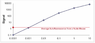
Pearl™ Imager系统使用了基于CCD、专门为近红外活体成像设计的光学系统。在近红外波段进行小动物活体成像时,组织的自发背景荧光和光散射比可见光区域更低,降低背景低的同时增加了检测灵敏度。而且,Pearl™ Imager系统的激发光源是近红外固态激光器,近红外激光器产生的激发光比白光具有更深的组织穿透性,即使更深层、更小的目标也能够检测到,因此,可以进行早期肿瘤检测。而且,成像的动力学范围宽,每个通道图像都具有6个数量级的动力学范围。

图示Pearl™ Imager系统非常宽的动态检测范围。IRDye800CW 染料0-10uM的浓度梯度递增成像,成像信号是目标区域总的荧光信号减去背景荧光信号。
简单、方便的操作

可分离的成像床能够快速将成像老鼠从预麻室转移到机器上进行成像,而且,成像床具有温控功能,维持成像老鼠的正常体温。
滑轮式成像抽屉,非常方便操作成像小鼠。
轻按成像床关闭按钮,然后再点击软件,无需设置曝光参数,就可以进行小动物成像。

完整解决方案
美国Li-COR公司的Pearl™ Imager小动物成像系统整合Odyssey双色红外激光成像系统组成体内和体外的多功能成像平台。该平台的体内成像或体外成像均使用该公司研发的、行业领先的IRDye染料,保证体内体外成像结果的可比性。目前,没有其他成像系统能够满足在体内和体外成像均使用同一种染料开展研究工作。

■ 红外光学试剂
肿瘤试剂
926-08446 IRDye® 800CW EGF Optical Probe
926-08946 IRDye 800CW 2-DG Optical Probe
926-09889 IRDye 800CW RGD Optical Probe
结构试剂
926-09375 IRDye® 800CW BoneTag™ (40 nmol)
926-09376 IRDye 680 BoneTag (80 nmol)
标记染料
929-70020 IRDye 800CW NHS Ester
929-80020 IRDye 800CW Maleimide
929-70010 IRDye 700DX NHS Ester
929-90010 CellVue Burgundy
929-90020 CellVue NIR815

■ 应用
肿瘤成像-IRDye 800CW EGF Optical Probe

许多肿瘤(肾细胞癌、非小细胞肺癌、神经胶质瘤、卵巢癌、膀胱癌、胰腺癌、结肠癌和乳腺癌等)的发生都会出现EGFR过量表达, IRDye 800CW EGF 探针是EGFR的定把探针,该标记探针和EGFR结合,可以示踪肿瘤的发生和转移等。

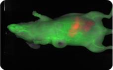
IRDye 800CW EGF示踪的是前列腺肿瘤(绿),IRDye 680 BoneTag探针表示骨骼结构(红)。
Imaging data taken usingLI-COR’s Pearl™ Imager。

肿瘤成像- IRDye 800CW 2-DG Optical Probe
肿瘤细胞的糖酵解过程一般会出现非正常的代谢旺盛。因此,可以用葡萄糖的类似物2-DG(2-deoxy-Dglucose)示踪肿瘤和肿瘤的转移。IRDye 800CW 2-DG(2-deoxy-Dglucose)光学探针被证实可以用来研究由细胞系A431(表皮癌), SW620(结肠癌), 3T3-L1, and PC3LMN4 引起的肿瘤。

Pearl™ Imager Data

肿瘤成像-IRDye800CW RGD Optical Probe
该探针由IRDye800CW的染料标记的环状五肽组成,识别基团RGD是一个三肽序列(Arginine, Glycine and Aspartic Acid)。RGD基团能特异结合细胞表面受体αvβ3 (细胞表面的异二聚体糖蛋白),可以研究和监测αvβ3受体过表达的相关疾病。该光学探针已经被证明能特异结合许多肿瘤细胞系。如U87, A431, PC3M-LN4和 22Rv1。
Data Courtesy of Dr. Kai Chen, StanfordUniversity

骨格结构成像-IRDye 680/800CW BoneTag™
IRDye 680/800CW BoneTag 是红外标记的钙螯合物的复合物,被证明在骨骼癌和骨骼结构方面具有非常有效的应用,能有效检测骨骼钙化、生长和形态学改变。
小分子药物成像
IRDye 800CW标记的小分子药物PEG能够结合血管壁,通过Pearl™ Imager小动物活体成像系统可以得到小鼠血管的清楚图像。

This data, courtesy of Dr. Kazuki Sugahara University of California, Santa Barbara

解剖器官成像

Data Courtesy of Dr. Dennis Wolff, CreightonUniversity


IRDye® 800CW BoneTag™ 光学探头,BrightSite™ IRDye® in vivo Imaging Agents,IRDye® 800CW EGF Optical Probe

BrightSite™  IRDye® in vivo  Imaging Agents

IRDye® 800CW 2-DG Optical Probe

BrightSite™  IRDye® in vivo  Imaging Agents

IRDye® 800CW EGF Optical Probe

BrightSite™  IRDye® in vivo  Imaging Agents

IRDye® 800CW RGD Optical Probe

BrightSite™  IRDye® in vivo  Imaging Agents

IRDye® 800CW BoneTag™ Optical Probe,IRDye® 800CW BoneTag™ 光学探头

BrightSite™  IRDye® in vivo  Imaging Agents

IRDye® 680RD BoneTag™ Optical Probe

BrightSite™  IRDye® in vivo  Imaging Agents

IRDye® 800CW PEG Fluorescent Contrast Agent

Every LI-COR BrightSite IRDye in vivo imaging agent has been carefully validated with cultured cell assays, microscopy, in vivo imaging of animal models, and histology to ensure high affinity and specificity.

BrightSite IRDye Small Animal Imaging Agents are:

  • Ready-to-use probes that are simple to administer, allowing you to begin animal studies immediately
  • Sensitive, offering the high signal-to-noise advantages of near-infrared fluorescence which are especially important for small or deep targets
  • Cited in several hundred peer-reviewed publications so you can use them with confidence
  • Imaged with any small animal imaging equipment with appropriate 680 nm or 800 nm filter sets
  • Compatible with most small animal imaging systems, including instruments from LI-COR Biosciences (Pearl® and Odyssey®), PerkinElmer (Xenogen, Caliper, CRi, and VisEn), and Bruker (Carestream and Kodak)
  • 易于管理的即用型探针,可让您立即开始动物研究
    灵敏,提供近红外荧光的高信噪比优势,这对于小目标或深目标尤其重要
    在数百篇同行评审的出版物中被引用,因此您可以放心使用它们
    使用任何带有适当 680 nm 或 800 nm 滤光片组的小动物成像设备进行成像
    与大多数小动物成像系统兼容,包括来自 LI-COR Biosciences(Pearl® 和 Odyssey®)、PerkinElmer(Xenogen、Caliper、CRi 和 VisEn)和 Bruker(Carestream 和 Kodak)的仪器

IRDye 800CW absorption/emission near 800 nm matches near-infrared absorption minima for bodily fluids and tissues, resulting in excellent tissue penetration, making it ideal for in vivo imaging.

BrightSite™  IRDye® in vivo  Imaging Agents
Molar extinction coefficient characteristics of water, hemoglobin and oxygenated hemoglobin (400-1000 nm). IRDye infrared dyes have ideal excitation/emission wavelengths for in vivo imaging.

Detect Apoptotic and Necrotic Cells, Bacteria and Other Anionic Membranes

LI-COR also offers the PSVue® 794 Reagent Kit which can be used to detect apoptotic and necrotic cells, bacteria and other anionic membranes.

LI-COR 还提供 PSVue® 794 试剂盒,可用于检测凋亡和坏死细胞、细菌和其他阴离子膜。

Fluorescently Label Cells or Tissues for in vivo or in vitro Studies

CellVue® fluorescent imaging kits use proprietary labeling technology to stably incorporate fluorescent dyes containing long aliphatic hydrocarbon tails into lipid membranes.1 They are useful for researchers working in all aspects of science and technology where fluorescently-labeled cells and/or tissues are required. CellVue dyes also provide researchers with valuable tools for many in vivo and in vitro cell studies using fluorescent membrane labels.

CellVue® 荧光成像试剂盒使用专有的标记技术,将含有长脂肪烃尾的荧光染料稳定地掺入脂质膜中。1 它们适用于在需要荧光标记的细胞和/或组织的科学和技术的各个方面工作的研究人员。 CellVue 染料还为研究人员提供了许多使用荧光膜标记进行体内和体外细胞研究的宝贵工具。

CellVue dyes consist of long aliphatic hydrocarbon tails linked to a polar fluorescent chromophore.

  • These extremely lipophilic fluorescent dyes rapidly and stably integrate into the phospholipid membrane of cells or other membrane-containing bioparticles by non-covalent interactions
  • The dyes are stably maintained within the lipid bilayer through strong hydrophobic interactions and do not transfer into the unstained membranes of adjacent cells, which permits a labeled cell to be tracked for extended periods of time
  • The rapid incorporation of the dye allows for immediate analysis of cell functions without a waiting period

BrightSite™  IRDye® in vivo  Imaging Agents

Molar extinction coefficient characteristics of water, hemoglobin and oxygenated hemoglobin (400-1000 nm). IRDye infrared dyes have ideal excitation/emission wavelengths for in vivo imaging.

Selecting the Right Optical Imaging Agent

Application IRDye EGF IRDye RGD IRDye 2-DG IRDye PEG IRDye BoneTag CellVue PSVue
Tumor Imaging BrightSite™  IRDye® in vivo  Imaging Agents BrightSite™  IRDye® in vivo  Imaging Agents BrightSite™  IRDye® in vivo  Imaging Agents BrightSite™  IRDye® in vivo  Imaging Agents
Metabolic Imaging BrightSite™  IRDye® in vivo  Imaging Agents
Inflammation/Arthritis BrightSite™  IRDye® in vivo  Imaging Agents BrightSite™  IRDye® in vivo  Imaging Agents
Vasculature (Contrast) BrightSite™  IRDye® in vivo  Imaging Agents
Lymphatic Imaging
Lymph Node Imaging
Structural Imaging BrightSite™  IRDye® in vivo  Imaging Agents
Cell Trafficking BrightSite™  IRDye® in vivo  Imaging Agents
Apoptosis BrightSite™  IRDye® in vivo  Imaging Agents

References

  1. Horan, P. K., and Slezak, S. E. (1989) Nature, 340. 167-168.