Accessories > Upper Buffer Tank Lid Assembly,上缓冲罐盖组件

MCE抑制剂∣Abcam CST Santa抗体∣蛋白、酶、磁珠
Accessories > Upper Buffer Tank Lid Assembly,上缓冲罐盖组件

Accessories > User-Applied Comb Support,用户应用的梳状支撑

This support bar is applied to rectangular combs to allow for appropriate well depth.
该支撑杆应用于矩形梳,以允许适当的井深度。
Reagents > Vinculin Rabbit Monoclonal Antibody for Normalization,用于标准化的 Vinculin 兔单克隆抗体

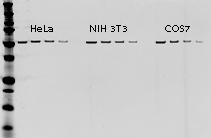
Vinculin is a highly conserved cytoskeletal protein. The Vinculin primary antibody can be used as an internal loading control for normalization and is particularly effective when detecting high molecular weight targets.
The expression of Vinculin, or any housekeeping protein (HKP), should be validated to ensure that its expression does not change under experimental conditions.
Once validated, Vinculin primary antibodies can be used for the detection of Vinculin when performing multiplex Western blot detection.
Detect Vinculin Rabbit Monoclonal Antibody with IRDye® Goat anti-Rabbit or IRDye Donkey anti-Rabbit secondary antibodies.
Other options for housekeeping protein normalization
Vinculin 是一种高度保守的细胞骨架蛋白。 Vinculin 一抗可用作标准化的内部上样对照,在检测高分子量目标时特别有效。
应验证 Vinculin 或任何管家蛋白 (HKP) 的表达,以确保其表达在实验条件下不会改变。
一旦通过验证,在进行多重蛋白质印迹检测时,Vinculin 一抗可用于检测 Vinculin。
使用 IRDye® Goat anti-Rabbit 或 IRDye Donkey anti-Rabbit 二抗检测 Vinculin Rabbit Monoclonal Antibody。
管家蛋白质标准化的其他选择
Vinculin antibody is supplied in 10 mM HEPES (pH 7.5), 150 mM NaCl, 100 µg/mL BSA, 50% glycerol, and <0.02% sodium azide.
Do not aliquot the antibody.
| Properties | Vinculin Rabbit Monoclonal Antibody (P/N 926-42215) |
|---|---|
| Species Cross-Reactivity | Human, mouse, rat, monkey |
| Target Molecular Weight | 124 kDa |
| Isotype | Rabbit IgG |
| Specificity/Sensitivity | Detects endogenous levels of total vinculin protein and metavinculin, a 145 kDa splice variant of vinculin |
| Immunogen | A recombinant protein that is specific to the amino terminus of human vinculin protein |
| Tested Application | Western blot (WB), Immunohistochemistry (IHC), Flow Cytometry (F) |

Reagents > VRDye™ 490 Goat anti-Mouse IgG Secondary Antibody,VRDye™ 490 山羊抗小鼠 IgG 二抗

Mouse IgG paraproteins
Isolation of specific antibodies was accomplished by affinity chromatography using pooled mouse IgG covalently linked to agarose. Based on ELISA and flow cytometry, this antibody reacts with the heavy and light chains of mouse IgG1, IgG2a, IgG2b, and IgG3, and with the light chains of mouse IgM and IgA. This antibody was tested by dot blot and and/or solid-phase adsorbed for minimal cross-reactivity with human, rabbit, goat, rat, and horse serum proteins, but may cross-react with immunoglobulins from other species. The conjugate has been specifically tested and qualified for immunofluorescence microscopy and flow cytometry applications.
使用与琼脂糖共价连接的混合小鼠 IgG 通过亲和层析实现特异性抗体的分离。 基于 ELISA 和流式细胞术,该抗体与小鼠 IgG1、IgG2a、IgG2b 和 IgG3 的重链和轻链以及小鼠 IgM 和 IgA 的轻链反应。 通过斑点印迹和/或固相吸附测试该抗体与人、兔、山羊、大鼠和马血清蛋白的交叉反应最小,但可能与来自其他物种的免疫球蛋白交叉反应。 该偶联物经过专门测试,可用于免疫荧光显微镜和流式细胞术应用。
VRDye 490 secondary antibodies are supplied as purified immunoglobulin conjugates, lyophilized in phosphate-buffered saline, pH 7.4. Protect from light. Store at 4 °C prior to reconstitution.
Each vial contains 10 mg/mL BSA (free of IgG and protease) as a stabilizer and 0.01% sodium azide as a preservative, after reconstitution. Concentration is 1.0 mg/mL when reconstituted as directed. Refer to the pack insert for details on reconstitution.
VRDye 490 二抗以纯化的免疫球蛋白偶联物形式提供,在磷酸盐缓冲盐水中冻干,pH 7.4。 避光。 在重组前储存在 4°C。
复溶后,每瓶含有 10 mg/mL BSA(不含 IgG 和蛋白酶)作为稳定剂和 0.01% 叠氮化钠作为防腐剂。 按照指示重新配制时,浓度为 1.0 mg/mL。 有关重组的详细信息,请参阅包装插页。
| Application | Recommended | Suggested Range |
|---|---|---|
| Immunofluorescence Microscopy | 1:400 | 1:100 – 1:1,000 |
| Flow Cytometry | 1:1,000 | 1:200 – 1:2,000 |
| Other | User-optimized |
Suggested working dilutions are given as a guide only. Optimum dilutions will vary and should be determined empirically.
Reagents > VRDye™ 490 Protein Labeling Kits,VRDye™ 490 蛋白质标记试剂盒

These kits may be used to label proteins spanning the molecular weight range of 50 kDa to 200 kDa. Labeled antibodies can be used for applications such as flow cytometry, microscopy, and immunohistochemistry.
这些试剂盒可用于标记分子量范围为 50 kDa 至 200 kDa 的蛋白质。 标记的抗体可用于流式细胞术、显微镜检查和免疫组织化学等应用。

This kit is optimized for labeling 1 mg of a primary or secondary IgG antibody.
This kit is optimized for labeling 100 µg of a primary or secondary IgG antibody.
该试剂盒针对标记 100 µg 一抗或二抗 IgG 进行了优化。
Reagents > VRDye™ 549 Goat anti-Mouse IgG Secondary Antibody,VRDye™ 549 山羊抗小鼠 IgG 二抗

Mouse IgG paraproteins
Isolation of specific antibodies was accomplished by affinity chromatography using pooled mouse IgG covalently linked to agarose. Based on ELISA and flow cytometry, this antibody reacts with the heavy and light chains of mouse IgG1, IgG2a, IgG2b, and IgG3, and with the light chains of mouse IgM and IgA. This antibody was tested by dot blot and and/or solid-phase adsorbed for minimal cross-reactivity with human, rabbit, goat, rat, and horse serum proteins, but may cross-react with immunoglobulins from other species. The conjugate has been specifically tested and qualified for immunofluorescence microscopy and flow cytometry applications.
使用与琼脂糖共价连接的混合小鼠 IgG 通过亲和层析实现特异性抗体的分离。 基于 ELISA 和流式细胞术,该抗体与小鼠 IgG1、IgG2a、IgG2b 和 IgG3 的重链和轻链以及小鼠 IgM 和 IgA 的轻链反应。 通过斑点印迹和/或固相吸附测试该抗体与人、兔、山羊、大鼠和马血清蛋白的交叉反应最小,但可能与来自其他物种的免疫球蛋白交叉反应。 该偶联物经过专门测试,可用于免疫荧光显微镜和流式细胞术应用。
VRDye 549 secondary antibodies are supplied as purified immunoglobulin conjugates, lyophilized in phosphate-buffered saline, pH 7.4. Protect from light. Store at 4 °C prior to reconstitution.
Each vial contains 10 mg/mL BSA (free of IgG and protease) as a stabilizer and 0.01% sodium azide as a preservative, after reconstitution. Concentration is 1.0 mg/mL when reconstituted as directed. Refer to the pack insert for details on reconstitution.
VRDye 549 二抗以纯化的免疫球蛋白偶联物形式提供,在 pH 7.4 的磷酸盐缓冲盐水中冻干。 避光。 在重组前储存在 4°C。
复溶后,每瓶含有 10 mg/mL BSA(不含 IgG 和蛋白酶)作为稳定剂和 0.01% 叠氮化钠作为防腐剂。 按照指示重新配制时,浓度为 1.0 mg/mL。 有关重组的详细信息,请参阅包装插页。
| Application | Recommended | Suggested Range |
|---|---|---|
| Immunofluorescence Microscopy | 1:400 | 1:100 – 1:1,000 |
| Flow Cytometry | 1:1,000 | 1:200 – 1:2,000 |
| Other | User optimized |
Suggested working dilutions are given as a guide only. Optimum dilutions will vary and should be determined empirically.
Reagents > VRDye™ 490 Donkey anti-Goat IgG Secondary Antibody,VRDye™ 490 驴抗山羊 IgG 二抗

Goat IgG
The antibody was isolated from antisera by immunoaffinity chromatography using antigens coupled to agarose beads. Based on ELISA, this antibody reacts with heavy and light chains of goat IgG and sheep IgG. This antibody was tested by dot blot and/or solid phase adsorbed for minimal cross-reactivity with human, mouse, rabbit, rat, chicken, guinea pig, hamster, swine, and horse serum proteins but may cross-react with immunoglobulins from other species. The conjugate has been specifically tested and qualified for Western blot applications.
使用与琼脂糖珠偶联的抗原,通过免疫亲和层析从抗血清中分离抗体。 基于 ELISA,该抗体与山羊 IgG 和绵羊 IgG 的重链和轻链反应。 该抗体通过斑点印迹和/或固相吸附测试,与人、小鼠、兔、大鼠、鸡、豚鼠、仓鼠、猪和马血清蛋白的交叉反应最小,但可能与来自其他物种的免疫球蛋白交叉反应 . 该偶联物经过专门测试,可用于蛋白质印迹应用。
VRDye 490 secondary antibodies are supplied as purified immunoglobulin conjugates, lyophilized in phosphate-buffered saline, pH 7.4. Protect from light. Store at 4 °C before and after reconstitution.
Each vial contains 10 mg/mL BSA (free of IgG and protease) as a stabilizer and 0.01% sodium azide as a preservative, after reconstitution. Concentration is 1.0 mg/mL when reconstituted as directed. Refer to the pack insert for details on reconstitution.
VRDye 490 二抗以纯化的免疫球蛋白偶联物形式提供,在磷酸盐缓冲盐水中冻干,pH 7.4。 避光。 在重组之前和之后储存在 4°C。
复溶后,每瓶含有 10 mg/mL BSA(不含 IgG 和蛋白酶)作为稳定剂和 0.01% 叠氮化钠作为防腐剂。 按照指示重新配制时,浓度为 1.0 mg/mL。 有关重组的详细信息,请参阅包装插页。
| Application | Recommended | Suggested Range |
|---|---|---|
| Western Blot Detection | 1:10,000 | 1:5,000 – 1:15,000 |
| Other | User-optimized |
Suggested working dilutions are given as a guide only. Optimum dilutions will vary and should be determined empirically.
Reagents > VRDye™ 490 Donkey anti-Mouse IgG Secondary Antibody,VRDye™ 490 驴抗小鼠 IgG 二抗

Mouse IgG
The antibody was isolated by affinity chromatography using antigens coupled to agarose beads. Based on ELISA, this antibody reacts with heavy and light chains of mouse IgG and the light chains of mouse IgM and IgA. This antibody was tested by ELISA and/or solid phase adsorbed to ensure minimal cross-reaction with bovine, chicken, goat, guinea pig, horse, human, rabbit, and sheep serum proteins but may cross-react with immunoglobulins from other species. The conjugate has been specifically tested and qualified for Western blot applications.
使用与琼脂糖珠偶联的抗原通过亲和层析分离抗体。 基于 ELISA,该抗体与小鼠 IgG 的重链和轻链以及小鼠 IgM 和 IgA 的轻链反应。 该抗体通过 ELISA 和/或固相吸附测试,以确保与牛、鸡、山羊、豚鼠、马、人、兔和绵羊血清蛋白的交叉反应最小,但可能与来自其他物种的免疫球蛋白交叉反应。 该偶联物经过专门测试,可用于蛋白质印迹应用。
VRDye 490 secondary antibodies are supplied as purified immunoglobulin conjugates, lyophilized in phosphate-buffered saline, pH 7.4. Protect from light. Store at 4 °C before and after reconstitution.
Each vial contains 10 mg/mL BSA (free of IgG and protease) as a stabilizer and 0.01% sodium azide as a preservative, after reconstitution. Concentration is 1.0 mg/mL when reconstituted as directed. Refer to the pack insert for details on reconstitution.
VRDye 490 二抗以纯化的免疫球蛋白偶联物形式提供,在磷酸盐缓冲盐水中冻干,pH 7.4。 避光。 在重组之前和之后储存在 4°C。
复溶后,每瓶含有 10 mg/mL BSA(不含 IgG 和蛋白酶)作为稳定剂和 0.01% 叠氮化钠作为防腐剂。 按照指示重新配制时,浓度为 1.0 mg/mL。 有关重组的详细信息,请参阅包装插页。
| Application | Recommended | Suggested Range |
|---|---|---|
| Western Blot Detection | 1:10,000 | 1:5,000 – 1:15,000 |
| Other | User-optimized |
Suggested working dilutions are given as a guide only. Optimum dilutions will vary and should be determined empirically.
Reagents > VRDye™ 490 Donkey anti-Rabbit IgG Secondary Antibody,VRDye™ 490 驴抗兔 IgG 二抗

Rabbit IgG
The antibody was isolated by affinity chromatography using antigens coupled to agarose beads. Based on ELISA, this antibody reacts with heavy and light chains of rabbit IgG and light chains common to most rabbit immunoglobulins. This antibody has been tested by ELISA and/or solid phase adsorbed to ensure minimal cross-reaction with bovine, chicken, goat, guinea pig, hamster, horse, human, mouse, rat, and sheep serum proteins but may cross-react with immunoglobulins from other species. The conjugate has been specifically tested and qualified for Western blot applications.
使用与琼脂糖珠偶联的抗原通过亲和层析分离抗体。 基于 ELISA,该抗体与兔 IgG 的重链和轻链以及大多数兔免疫球蛋白常见的轻链发生反应。 该抗体已通过 ELISA 和/或固相吸附测试,以确保与牛、鸡、山羊、豚鼠、仓鼠、马、人、小鼠、大鼠和绵羊血清蛋白的交叉反应最小,但可能与免疫球蛋白发生交叉反应 来自其他物种。 该偶联物经过专门测试,可用于蛋白质印迹应用。
VRDye 490 secondary antibodies are supplied as purified immunoglobulin conjugates, lyophilized in phosphate-buffered saline, pH 7.4. Protect from light. Store at 4 °C before and after reconstitution.
Each vial contains 10 mg/mL BSA (free of IgG and protease) as a stabilizer and 0.01% sodium azide as a preservative, after reconstitution. Concentration is 1.0 mg/mL when reconstituted as directed. Refer to the pack insert for details on reconstitution.
VRDye 490 二抗以纯化的免疫球蛋白偶联物形式提供,在磷酸盐缓冲盐水中冻干,pH 7.4。 避光。 在重组之前和之后储存在 4°C。
复溶后,每瓶含有 10 mg/mL BSA(不含 IgG 和蛋白酶)作为稳定剂和 0.01% 叠氮化钠作为防腐剂。 按照指示重新配制时,浓度为 1.0 mg/mL。 有关重组的详细信息,请参阅包装插页。
| Application | Recommended | Suggested Range |
|---|---|---|
| Western Blot Detection | 1:10,000 | 1:5,000 – 1:15,000 |
| Other | User-optimized |
Suggested working dilutions are given as a guide only. Optimum dilutions will vary and should be determined empirically.
Reagents > VRDye™ 490 Goat anti-Rabbit IgG Secondary Antibody,VRDye™ 490 山羊抗兔 IgG 二抗

Rabbit IgG
Isolation of specific antibodies was accomplished by affinity chromatography using pooled rabbit IgG covalently linked to agarose. Based on ELISA and flow cytometry, this antibody reacts with the heavy and light chains of rabbit IgG, and with the light chains of rabbit IgM and IgA. This antibody was tested by dot blot and and/or solid-phase adsorbed for minimal cross-reactivity with human, mouse, rat, sheep, and chicken serum proteins, but may cross-react with immunoglobulins from other species. The conjugate has been specifically tested and qualified for immunofluorescence microscopy and flow cytometry applications.
使用与琼脂糖共价连接的混合兔 IgG,通过亲和层析实现特异性抗体的分离。 基于 ELISA 和流式细胞术,该抗体与兔 IgG 的重链和轻链以及兔 IgM 和 IgA 的轻链反应。 通过斑点印迹和/或固相吸附测试该抗体与人、小鼠、大鼠、绵羊和鸡血清蛋白的交叉反应最小,但可能与来自其他物种的免疫球蛋白交叉反应。 该偶联物经过专门测试,可用于免疫荧光显微镜和流式细胞术应用。
VRDye 490 secondary antibodies are supplied as purified immunoglobulin conjugates, lyophilized in phosphate-buffered saline, pH 7.4. Protect from light. Store at 4 °C prior to reconstitution.
Each vial contains 10 mg/mL BSA (free of IgG and protease) as a stabilizer and 0.01% sodium azide as a preservative, after reconstitution. Concentration is 1.0 mg/mL when reconstituted as directed. Refer to the pack insert for details on reconstitution.
VRDye 490 二抗以纯化的免疫球蛋白偶联物形式提供,在磷酸盐缓冲盐水中冻干,pH 7.4。 避光。 在重组前储存在 4°C。
复溶后,每瓶含有 10 mg/mL BSA(不含 IgG 和蛋白酶)作为稳定剂和 0.01% 叠氮化钠作为防腐剂。 按照指示重新配制时,浓度为 1.0 mg/mL。 有关重组的详细信息,请参阅包装插页。
| Application | Recommended | Suggested Range |
|---|---|---|
| Immunofluorescence Microscopy | 1:400 | 1:100 – 1:1,000 |
| Flow Cytometry | 1:1,000 | 1:200 – 1:2,000 |
| Other | User-optimized |
Suggested working dilutions are given as a guide only. Optimum dilutions will vary and should be determined empirically.

Reagents > VRDye™ 490 Streptavidin,VRDye™ 490 链霉亲和素

VRDye 490 Streptavidin is supplied as a liquid in buffer containing 10 mM phosphate, 183 mM NaCl, 2.7 mM KCl, pH 7.4 with sodium azide 0.005% (w/v) as a preservative.
To use, centrifuge briefly before use to eliminate aggregates that may have formed in solution. This will reduce non-specific background staining.
For membrane-based applications and In-Gel Westerns, it is recommended to add SDS (0.02% to 0.1% final concentration), in addition to Tween® 20 (0.1 to 0.2% final concentration) during the detection incubation step to reduce non-specific background staining.
VRDye 490 链霉亲和素以液体形式提供,缓冲液中含有 10 mM 磷酸盐、183 mM NaCl、2.7 mM KCl,pH 7.4,叠氮化钠 0.005% (w/v) 作为防腐剂。
要使用,请在使用前短暂离心以消除可能在溶液中形成的聚集体。 这将减少非特异性背景染色。
对于基于膜的应用和 In-Gel Western,建议在检测孵育步骤中添加 SDS(0.02% 至 0.1% 最终浓度)和 Tween® 20(0.1% 至 0.2% 最终浓度)以减少非 特定背景染色。
| Application | Suggested Range | Tween 20* | SDS* |
|---|---|---|---|
| Western blot detection | 1:2,000 – 1:5,000 | 0.1 – 0.2% (v/v) | 0.02 – 0.1% (v/v) |
| Other | User optimized | User optimized | User optimized |
Optimum dilutions will vary and should be determined empirically.
* Added to reduce non-specific background staining.
Reagents > VRDye™ 549 Donkey anti-Goat IgG Secondary Antibody,VRDye™ 549 驴抗山羊 IgG 二抗

Goat IgG
The antibody was isolated from antisera by immunoaffinity chromatography using antigens coupled to agarose beads. Based on ELISA, this antibody reacts with heavy and light chains of goat IgG and sheep IgG. This antibody was tested by dot blot and/or solid phase adsorbed for minimal cross-reactivity with human, mouse, rabbit, rat, chicken, guinea pig, hamster, swine, and horse serum proteins but may cross-react with immunoglobulins from other species. The conjugate has been specifically tested and qualified for Western blot applications.
使用与琼脂糖珠偶联的抗原,通过免疫亲和层析从抗血清中分离抗体。 基于 ELISA,该抗体与山羊 IgG 和绵羊 IgG 的重链和轻链反应。 该抗体通过斑点印迹和/或固相吸附测试,与人、小鼠、兔、大鼠、鸡、豚鼠、仓鼠、猪和马血清蛋白的交叉反应最小,但可能与来自其他物种的免疫球蛋白交叉反应 . 该偶联物经过专门测试,可用于蛋白质印迹应用。
VRDye 549 secondary antibodies are supplied as purified immunoglobulin conjugates, lyophilized in phosphate-buffered saline, pH 7.4. Protect from light. Store at 4 °C before and after reconstitution.
Each vial contains 10 mg/mL BSA (free of IgG and protease) as a stabilizer and 0.01% sodium azide as a preservative, after reconstitution. Concentration is 1.0 mg/mL when reconstituted as directed. Refer to the pack insert for details on reconstitution.
VRDye 549 二抗以纯化的免疫球蛋白偶联物形式提供,在 pH 7.4 的磷酸盐缓冲盐水中冻干。 避光。 在重组之前和之后储存在 4°C。
复溶后,每瓶含有 10 mg/mL BSA(不含 IgG 和蛋白酶)作为稳定剂和 0.01% 叠氮化钠作为防腐剂。 按照指示重新配制时,浓度为 1.0 mg/mL。 有关重组的详细信息,请参阅包装插页。
| Application | Recommended | Suggested Range |
|---|---|---|
| Western Blot Detection | 1:10,000 | 1:5,000 – 1:15,000 |
| Other | User-optimized |
Suggested working dilutions are given as a guide only. Optimum dilutions will vary and should be determined empirically.
Reagents > VRDye™ 549 Donkey anti-Mouse IgG Secondary Antibody,VRDye™ 549 驴抗小鼠 IgG 二抗

Mouse IgG
The antibody was isolated by affinity chromatography using antigens coupled to agarose beads. Based on ELISA, this antibody reacts with heavy and light chains of mouse IgG and the light chains of mouse IgM and IgA. This antibody was tested by ELISA and/or solid phase adsorbed to ensure minimal cross-reaction with bovine, chicken, goat, guinea pig, horse, human, rabbit, and sheep serum proteins but may cross-react with immunoglobulins from other species. The conjugate has been specifically tested and qualified for Western blot applications.
使用与琼脂糖珠偶联的抗原通过亲和层析分离抗体。 基于 ELISA,该抗体与小鼠 IgG 的重链和轻链以及小鼠 IgM 和 IgA 的轻链反应。 该抗体通过 ELISA 和/或固相吸附测试,以确保与牛、鸡、山羊、豚鼠、马、人、兔和绵羊血清蛋白的交叉反应最小,但可能与来自其他物种的免疫球蛋白交叉反应。 该偶联物经过专门测试,可用于蛋白质印迹应用。
VRDye 549 secondary antibodies are supplied as purified immunoglobulin conjugates, lyophilized in phosphate-buffered saline, pH 7.4. Protect from light. Store at 4 °C before and after reconstitution.
Each vial contains 10 mg/mL BSA (free of IgG and protease) as a stabilizer and 0.01% sodium azide as a preservative, after reconstitution. Concentration is 1.0 mg/mL when reconstituted as directed. Refer to the pack insert for details on reconstitution.
VRDye 549 二抗以纯化的免疫球蛋白偶联物形式提供,在 pH 7.4 的磷酸盐缓冲盐水中冻干。 避光。 在重组之前和之后储存在 4°C。
复溶后,每瓶含有 10 mg/mL BSA(不含 IgG 和蛋白酶)作为稳定剂和 0.01% 叠氮化钠作为防腐剂。 按照指示重新配制时,浓度为 1.0 mg/mL。 有关重组的详细信息,请参阅包装插页。
| Application | Recommended | Suggested Range |
|---|---|---|
| Western Blot Detection | 1:10,000 | 1:5,000 – 1:15,000 |
| Other | User-optimized |
Suggested working dilutions are given as a guide only. Optimum dilutions will vary and should be determined empirically.
Reagents > VRDye™ 549 Donkey anti-Rabbit IgG Secondary Antibody,VRDye™ 549 驴抗兔 IgG 二抗

Rabbit IgG
The antibody was isolated by affinity chromatography using antigens coupled to agarose beads. Based on ELISA, this antibody reacts with heavy and light chains of rabbit IgG and light chains common to most rabbit immunoglobulins. This antibody has been tested by ELISA and/or solid phase adsorbed to ensure minimal cross-reaction with bovine, chicken, goat, guinea pig, hamster, horse, human, mouse, rat, and sheep serum proteins but may cross-react with immunoglobulins from other species. The conjugate has been specifically tested and qualified for Western blot applications.
使用与琼脂糖珠偶联的抗原通过亲和层析分离抗体。 基于 ELISA,该抗体与兔 IgG 的重链和轻链以及大多数兔免疫球蛋白常见的轻链发生反应。 该抗体已通过 ELISA 和/或固相吸附测试,以确保与牛、鸡、山羊、豚鼠、仓鼠、马、人、小鼠、大鼠和绵羊血清蛋白的交叉反应最小,但可能与免疫球蛋白发生交叉反应 来自其他物种。 该偶联物经过专门测试,可用于蛋白质印迹应用。
VRDye 549 secondary antibodies are supplied as purified immunoglobulin conjugates, lyophilized in phosphate-buffered saline, pH 7.4. Protect from light. Store at 4 °C before and after reconstitution.
Each vial contains 10 mg/mL BSA (free of IgG and protease) as a stabilizer and 0.01% sodium azide as a preservative, after reconstitution. Concentration is 1.0 mg/mL when reconstituted as directed. Refer to the pack insert for details on reconstitution.
VRDye 549 二抗以纯化的免疫球蛋白偶联物形式提供,在 pH 7.4 的磷酸盐缓冲盐水中冻干。 避光。 在重组之前和之后储存在 4°C。
复溶后,每瓶含有 10 mg/mL BSA(不含 IgG 和蛋白酶)作为稳定剂和 0.01% 叠氮化钠作为防腐剂。 按照指示重新配制时,浓度为 1.0 mg/mL。 有关重组的详细信息,请参阅包装插页。
| Application | Recommended | Suggested Range |
|---|---|---|
| Western Blot Detection | 1:10,000 | 1:5,000 – 1:15,000 |
| Other | User-optimized |
Suggested working dilutions are given as a guide only. Optimum dilutions will vary and should be determined empirically.
Reagents > VRDye™ 549 Goat anti-Rabbit IgG Secondary Antibody,VRDye™ 549 山羊抗兔 IgG 二抗

Rabbit IgG
Isolation of specific antibodies was accomplished by affinity chromatography using pooled rabbit IgG covalently linked to agarose. Based on ELISA and flow cytometry, this antibody reacts with the heavy and light chains of rabbit IgG, and with the light chains of rabbit IgM and IgA. This antibody was tested by dot blot and and/or solid-phase adsorbed for minimal cross-reactivity with human, mouse, rat, sheep, and chicken serum proteins, but may cross-react with immunoglobulins from other species. The conjugate has been specifically tested and qualified for immunofluorescence microscopy and flow cytometry applications.
使用与琼脂糖共价连接的混合兔 IgG,通过亲和层析实现特异性抗体的分离。 基于 ELISA 和流式细胞术,该抗体与兔 IgG 的重链和轻链以及兔 IgM 和 IgA 的轻链反应。 通过斑点印迹和/或固相吸附测试该抗体与人、小鼠、大鼠、绵羊和鸡血清蛋白的交叉反应最小,但可能与来自其他物种的免疫球蛋白交叉反应。 该偶联物经过专门测试,可用于免疫荧光显微镜和流式细胞术应用。
VRDye 549 secondary antibodies are supplied as purified immunoglobulin conjugates, lyophilized in phosphate-buffered saline, pH 7.4. Protect from light. Store at 4 °C prior to reconstitution.
Each vial contains 10 mg/mL BSA (free of IgG and protease) as a stabilizer and 0.01% sodium azide as a preservative, after reconstitution. Concentration is 1.0 mg/mL when reconstituted as directed. Refer to the pack insert for details on reconstitution.
VRDye 549 二抗以纯化的免疫球蛋白偶联物形式提供,在 pH 7.4 的磷酸盐缓冲盐水中冻干。 避光。 在重组前储存在 4°C。
复溶后,每瓶含有 10 mg/mL BSA(不含 IgG 和蛋白酶)作为稳定剂和 0.01% 叠氮化钠作为防腐剂。 按照指示重新配制时,浓度为 1.0 mg/mL。 有关重组的详细信息,请参阅包装插页。
| Application | Recommended | Suggested Range |
|---|---|---|
| Immunofluorescence Microscopy | 1:400 | 1:100 – 1:1,000 |
| Flow Cytometry | 1:1,000 | 1:200 – 1:2,000 |
| Other | User-optimized |
Suggested working dilutions are given as a guide only. Optimum dilutions will vary and should be determined empirically.

Reagents > VRDye™ 549 Streptavidin,VRDye™ 549 链霉亲和素

VRDye 549 Streptavidin is supplied as a liquid in buffer containing 10 mM phosphate, 183 mM NaCl, 2.7 mM KCl, pH 7.4 with sodium azide 0.005% (w/v) as a preservative.
To use, centrifuge briefly before use to eliminate aggregates that may have formed in solution. This will reduce non-specific background staining.
For membrane-based applications and In-Gel Westerns, it is recommended to add SDS (0.02% to 0.1% final concentration), in addition to Tween® 20 (0.1 to 0.2% final concentration) during the detection incubation step to reduce non-specific background staining.
VRDye 549 链霉亲和素以液体形式提供,缓冲液中含有 10 mM 磷酸盐、183 mM NaCl、2.7 mM KCl,pH 7.4,叠氮化钠 0.005% (w/v) 作为防腐剂。
要使用,请在使用前短暂离心以消除可能在溶液中形成的聚集体。 这将减少非特异性背景染色。
对于基于膜的应用和 In-Gel Western,建议在检测孵育步骤中添加 SDS(0.02% 至 0.1% 最终浓度)和 Tween® 20(0.1% 至 0.2% 最终浓度)以减少非 特定背景染色。
| Application | Suggested Range | Tween 20* | SDS* |
|---|---|---|---|
| Western blot detection | 1:2,000 – 1:5,000 | 0.1 – 0.2% (v/v) | 0.02 – 0.1% (v/v) |
| Other | User optimized | User optimized | User optimized |
Optimum dilutions will vary and should be determined empirically.
* Added to reduce non-specific background staining.
Reagents > VRDye™ 549 Protein Labeling Kits,VRDye™ 549 蛋白质标记试剂盒

These kits may be used to label proteins spanning the molecular weight range of 50 kDa to 200 kDa. Labeled antibodies can be used for applications such as flow cytometry, microscopy, and immunohistochemistry.
这些试剂盒可用于标记分子量范围为 50 kDa 至 200 kDa 的蛋白质。 标记的抗体可用于流式细胞术、显微镜检查和免疫组织化学等应用。

This kit is optimized for labeling 1 mg of a primary or secondary IgG antibody.
该试剂盒针对标记 1 mg 一抗或二抗 IgG 进行了优化。
This kit is optimized for labeling 100 µg of a primary or secondary IgG antibody.
Accessories > Western Blot Storage Bags,蛋白质印迹存储袋
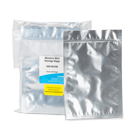
Western Blot Storage Bags are ideal for long-term storage of near-infrared fluorescent Western blot membranes. These bags protect blotted membranes from light, scratching, and photobleaching of fluorophores for later reprobing (detection of different targets), imaging, and documentation purposes.
Both nitrocellulose and PVDF membranes treated with primary and secondary antibodies can be stored dry for later imaging and documentation.
For blots that are being stored for reprobing, it is recommended that membranes be stripped first, and dried prior to storage.
蛋白质印迹存储袋是长期存储近红外荧光蛋白质印迹膜的理想选择。 这些袋子保护印迹膜免受光照、刮擦和荧光团的光漂白,以便以后重新探测(检测不同目标)、成像和记录目的。
用一抗和二抗处理的硝酸纤维素膜和 PVDF 膜都可以干燥储存,以供以后成像和记录。
对于储存用于重新检测的印迹,建议先剥离膜,并在储存前干燥。
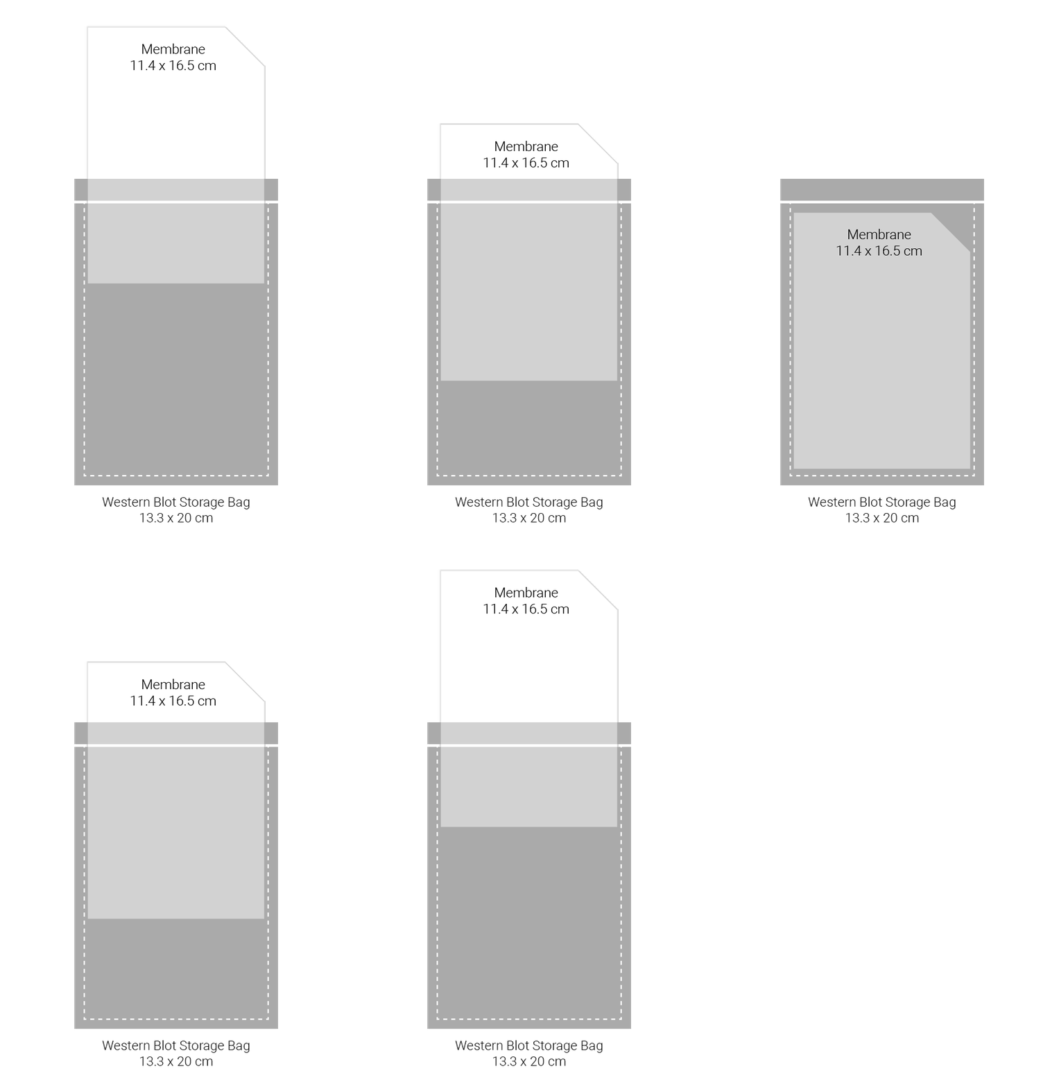
Each pack contains 40 resealable foil pouches (13.3 x 20 cm) for convenient storage of 11.4 x 16.5 cm membranes.
每包包含 40 个可重新密封的铝箔袋(13.3 x 20 厘米),方便存放 11.4 x 16.5 厘米的薄膜。
Reagents > Western Blotting Kit I RD (PBS/PVDF),蛋白质印迹试剂盒 I RD (PBS/PVDF)

See the full line of Odyssey Western Blotting Kits.查看完整的 Odyssey Western Blotting Kit 系列。
Reagents > Western Blotting Kit II RD (PBS/PVDF),蛋白质印迹试剂盒 II RD (PBS/PVDF)

See the full line of Western Blotting Kits.
Reagents > Western Blotting Kit III RD (PBS/NITRO),蛋白质印迹试剂盒 III RD (PBS/NITRO)

See the full line of Western Blotting Kits.
Reagents > Western Blotting Kit IV RD (PBS/NITRO),蛋白质印迹试剂盒 IV RD (PBS/NITRO)

See the full line of Western Blotting Kits.
Reagents > Western Blotting Kit V RD (TBS/PVDF),蛋白质印迹试剂盒 V RD (TBS/PVDF)
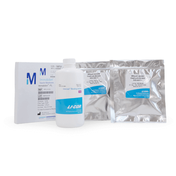
See the full line of Western Blotting Kits.
Reagents > Western Blotting Kit VI RD (TBS/PVDF),蛋白质印迹试剂盒 VI RD (TBS/PVDF)

See the full line of Western Blotting Kits.
Reagents > Western Blotting Kit VII RD (TBS/NITRO),蛋白质印迹试剂盒 VII RD (TBS/NITRO)

See the full line of Western Blotting Kits.
Reagents > Western Blotting Kit VIII RD (TBS/NITRO),蛋白质印迹试剂盒 VIII RD (TBS/NITRO)

See the full line of Western Blotting Kits.
Software > Western Key (Lite/C-DiGit®),西键 (Lite/C-DiGit®)

This is an optional key available for purchase separately with the following software:
With this key, you can analyze two-color Western blot and multiplex Western blot images using advanced analysis tools for normalization, sizing, and quantitation.
Note: The C-DiGit Blot Scanner cannot image two-color Western blots, but the software can import images acquired with other LI-COR instruments for analysis.
这是一个可选密钥,可与以下软件一起单独购买:
用于 C-DiGit® 印迹扫描仪的 Image Studio™ 软件
Image Studio 精简版软件
使用此密钥,您可以使用高级分析工具分析双色蛋白质印迹和多重蛋白质印迹图像,以进行标准化、大小调整和定量。
注意:C-DiGit Blot Scanner 无法对双色 Western blot 进行成像,但该软件可以导入使用其他 LI-COR 仪器采集的图像进行分析。
Reagents > WesternSure® Pre-stained Chemiluminescent Protein Ladder,WesternSure® 预染色化学发光蛋白阶梯

Western blot workflows with chemiluminescent protein ladders are prone to complications:
Note: The WesternSure Ladder is not recommended for use when stripping and reprobing Western blots. Stripping buffer alters the chemiluminescent functionality of the ladder, resulting in weak or no signal.
带有化学发光蛋白梯的蛋白质印迹工作流程容易出现并发症:
您的梯子可能需要带链球菌标记的 HRP
您的梯子可能依赖于 HRP 二抗检测到的 IgG 结合位点
可能需要反复试验来优化梯子的装载量
注意:剥离和重新检测蛋白质印迹时不建议使用 WesternSure Ladder。 剥离缓冲液会改变梯子的化学发光功能,导致信号微弱或无信号。
The WesternSure chemiluminescent protein ladder is the only pre-stained, multi-colored protein ladder for both film and digital chemiluminescence detection.
Not sure which protein ladder to choose? Learn more about how to choose the right protein ladder.
WesternSure 化学发光蛋白分子量标准是唯一一种用于胶片和数字化学发光检测的预染色、多色蛋白分子量标准。
适合您当前的协议,无需优化
不依赖链球菌标记的 HRP 或二抗结合
适用于各种基材
提供充满活力的配色方案,可让您快速定位膜
不确定选择哪种蛋白质阶梯? 详细了解如何选择正确的蛋白质阶梯。

Reagents > WesternSure® ECL Stripping Buffer for Chemiluminescent Westerns,用于化学发光西部片的 WesternSure® ECL 剥离缓冲液

WesternSure ECL Stripping Buffer is ideal for stripping and reprobing chemiluminescent Western blots. This Western blot stripping buffer:
Stripping buffers are not recommended if bands need to be quantified.
WesternSure ECL 剥离缓冲液是剥离和重新检测化学发光蛋白质印迹的理想选择。 这种蛋白质印迹剥离缓冲液:
从 PVDF 或硝酸纤维素膜上去除一抗和二抗
保持目标抗原的完整性,以实现高效的重新探测
与许多其他剥离缓冲液不同,不需要危险运输
如果需要量化条带,不建议使用剥离缓冲液。
注意:WesternSure ECL 剥离缓冲液不推荐用于超过 30 µg 的上样量。 当以 2X 或 5X 浓度使用时,它会表现出性能下降,并且加热溶液不会提高剥离效率。
WesternSure ECL Stripping Buffer is provided as a 5X concentrated solution. It is stored at room temperature.

Reagents > WesternSure® Goat anti-Rabbit HRP Secondary Antibody,WesternSure® 山羊抗兔 HRP 二抗
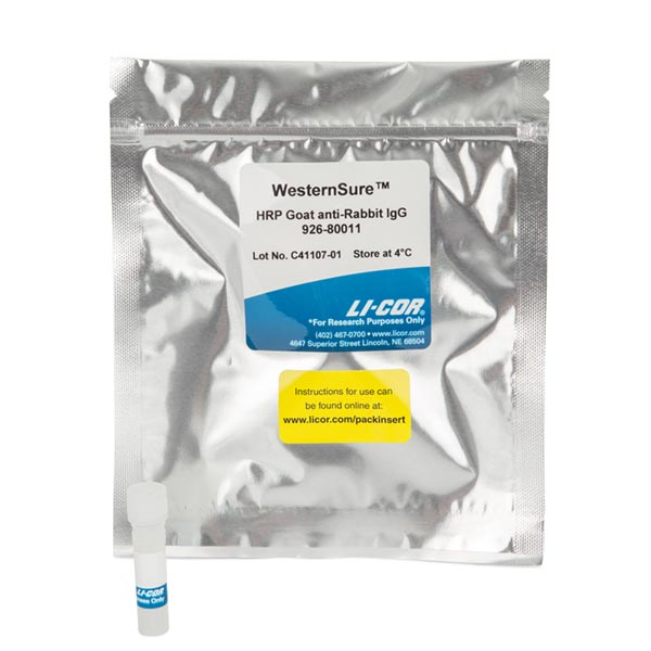
Rabbit IgG
The antibody was purified by affinity chromatography on pooled rabbit IgG covalently linked to agarose. Based on ELISA and/or flow cytometry, the antibody reacts with the heavy and light chains of rabbit IgG, and with the light chains of rabbit IgM. The antibody has minimal cross-reactivity with human and mouse immunoglobulins.
The antibody is supplied as purified immunoglobulin, supplied in 50% glycerol and 50% PBS, pH 7.4. No preservative has been added. Store at 4 °C.
| Substrate | Recommended Dilution Range |
|---|---|
| WesternSure PREMIUM Substrate | 1:5,000 – 1:100,000 |
| Other | User optimized |
Reagents > WesternSure® Pen
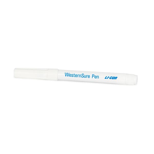
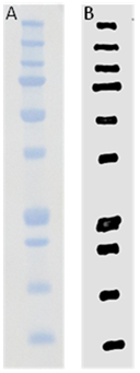
The WesternSure Pen is used to annotate visible protein ladders (such as Chameleon® Vue, P/N 928-50000) prior to chemiluminescent Western blot detection.
The pen is optimized for detection using the C-DiGit® Blot Scanner or the Odyssey® Fc Imaging System, and is also suitable for use with film or other imaging systems. This unique marker delivers an ink which emits light when incubated with commonly-used chemiluminescent substrates, including WesternSure PREMIUM Chemiluminescent Substrate. The ink is faintly visible for easy identification of marked membranes.
WesternSure Pen 用于在化学发光蛋白质印迹检测之前注释可见蛋白质梯(例如 Chameleon® Vue,P/N 928-50000)。
该笔针对使用 C-DiGit® 印迹扫描仪或 Odyssey® Fc 成像系统进行检测进行了优化,也适用于胶片或其他成像系统。 这种独特的标记提供的墨水在与常用的化学发光底物(包括 WesternSure PREMIUM 化学发光底物)一起孵育时会发光。 墨水隐约可见,便于识别标记的膜。
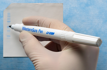
“It is so nice to be able to just draw right on the membrane and then see it.”
Reagents > WesternSure® PREMIUM Chemiluminescent Substrate,WesternSure® PREMIUM 化学发光底物

WesternSure PREMIUM Chemiluminescent Substrate is a highly sensitive, enhanced substrate for detecting horseradish peroxidase (HRP) on immunoblots.
The substrate pack contains Luminol Enhancer Solution and Stable Peroxidase Solution.
WesternSure PREMIUM 化学发光底物是一种高度灵敏的增强底物,用于检测免疫印迹上的辣根过氧化物酶 (HRP)。
孵育后 60 分钟以上的灵敏度与 SuperSignal® West Dura 相当
持续时间最长的最强信号
非常适合与数字成像系统一起使用
底物包包含鲁米诺增强剂溶液和稳定的过氧化物酶溶液。


Reagents > WesternSure® Goat anti-Mouse HRP Secondary Antibody,WesternSure® 山羊抗小鼠 HRP 二抗

Mouse IgG paraproteins
The antibody was purified by affinity chromatography on pooled mouse IgG covalently linked to agarose. Based on ELISA and/or flow cytometry, the antibody reacts with the heavy and light chains of mouse IgG1, IgG2a, IgG2b, and IgG3, along with the light chains of mouse IgM and IgA. The antibody has minimal cross-reactivity with human immunoglobulins.
The antibody is supplied as purified immunoglobulin, supplied in 50% glycerol and 50% PBS, pH 7.4. No preservative has been added. Store at 4 °C.
抗体以纯化的免疫球蛋白形式提供,在 50% 甘油和 50% PBS,pH 7.4 中提供。 没有添加防腐剂。 储存在 4°C。
| Substrate | Recommended Dilution Range |
|---|---|
| WesternSure PREMIUM Substrate | 1:5,000 – 1:100,000 |
| Other | User optimized |
Reagents > α-Tubulin Mouse Monoclonal Antibody for Normalization,用于标准化的 α-微管蛋白小鼠单克隆抗体

α-Tubulin primary antibody can be used as an internal loading control (ILC) for normalization.
The expression of α-tubulin, or any housekeeping protein (HKP), should be validated to ensure that its expression does not change under experimental conditions.
Once validated, α-tubulin primary antibodies can be used for the detection of α-tubulin when performing two-color detection.
Detect α-Tubulin Mouse Monoclonal Antibody with IRDye® Goat anti-Mouse or IRDye Donkey anti-Mouse secondary antibodies.
Other options for housekeeping protein normalization
α-微管蛋白一抗可用作内部上样对照 (ILC) 以进行标准化。
应验证 α-微管蛋白或任何管家蛋白 (HKP) 的表达,以确保其表达在实验条件下不会改变。
经验证后,α-微管蛋白一抗可在进行双色检测时用于检测 α-微管蛋白。
使用 IRDye® Goat anti-Mouse 或 IRDye Donkey anti-Mouse 二抗检测 α-Tubulin Mouse Monoclonal Antibody。
管家蛋白质标准化的其他选择
α-Tubulin antibody is supplied in 10 mM HEPES (pH 7.5), 150 mM NaCl, 100 µg/mL BSA, 50% glycerol, and <0.02% sodium azide.Do not aliquot the antibody.
α-微管蛋白抗体以 10 mM HEPES (pH 7.5)、150 mM NaCl、100 µg/mL BSA、50% 甘油和 <0.02% 叠氮化钠的形式提供。
不要等分抗体。
| Properties | α-Tubulin Mouse Monoclonal Antibody (P/N 926-42213) |
|---|---|
| Species Cross-Reactivity | Human, mouse, rat, monkey |
| Target Molecular Weight | 52 kDa |
| Isotype | Mouse IgG1 |
| Specificity/Sensitivity | Detects endogenous levels of total α-tubulin protein. α/β-tubulin heterodimers form the tubulin subunit common to all eukaryotic cells.
检测总 α-微管蛋白的内源水平。 α/β-微管蛋白异二聚体形成所有真核细胞共有的微管蛋白亚基。 |
| Immunogen | Full-length chicken α-tubulin (purified from brain extracts) |
| Tested Applications | Western blot (WB), Immunohistochemistry (IHC), Immunofluorescence (IF), Flow Cytometry (F)
蛋白质印迹 (WB)、免疫组织化学 (IHC)、免疫荧光 (IF)、流式细胞术 (F) |
Reagents > β-Actin Mouse Monoclonal Antibody for Normalization,用于标准化的 β-肌动蛋白小鼠单克隆抗体

The β-actin primary antibody can be used as an internal loading control (ILC) for normalization.
The expression of β-actin, or any housekeeping protein (HKP), should be validated to ensure that its expression does not change under experimental conditions.
Once validated, β-actin primary antibodies can be used for the detection of β-actin when performing two-color detection.
Detect β-Actin Mouse Primary Antibody with IRDye® Goat anti-Mouse or IRDye Donkey anti-Mouse secondary antibodies.
Other options for housekeeping protein normalization
β-肌动蛋白一抗可用作内部上样对照 (ILC) 以进行标准化。
应验证 β-肌动蛋白或任何管家蛋白 (HKP) 的表达,以确保其表达在实验条件下不会改变。
经验证后,β-actin 一抗可用于进行双色检测时对 β-actin 的检测。
使用 IRDye® Goat anti-Mouse 或 IRDye Donkey anti-Mouse 二抗检测 β-Actin Mouse Primary Antibody。
管家蛋白质标准化的其他选择
β-Actin antibody is supplied in 10 mM HEPES (pH 7.5), 150 mM NaCl, 100 µg/mL BSA, 50% glycerol, and <0.02% sodium azide.
Do not aliquot the antibody.
β-肌动蛋白抗体以 10 mM HEPES (pH 7.5)、150 mM NaCl、100 µg/mL BSA、50% 甘油和 <0.02% 叠氮化钠的形式提供。
不要等分抗体。
| Properties | β-Actin Mouse Monoclonal Antibody (P/N 926-42212) |
|---|---|
| Species Cross-Reactivity | Human, mouse, rat, monkey, hamster |
| Target Molecular Weight | 45 kDa |
| Isotype | Mouse IgG2b |
| Specificity/Sensitivity | Detects endogenous levels of β-actin protein |
| Immunogen | A synthetic peptide (KLH-coupled) that corresponds to the residues near the amino-terminus of human β-actin |
| Tested Applications | Western blot (WB), Immunohistochemistry (IHC), Immunofluorescence (IF), Flow Cytometry (F) |

Reagents > β-Actin Rabbit Monoclonal Antibody for Normalization,用于标准化的 β-肌动蛋白兔单克隆抗体,P/N 926-42210
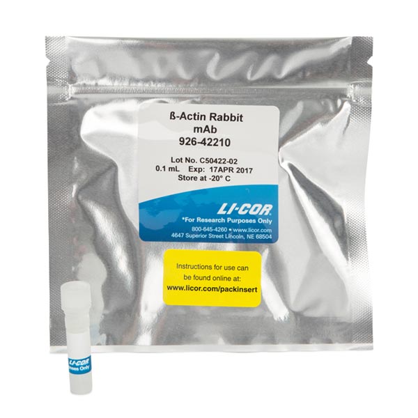
The β-actin primary antibody can be used as an internal loading control (ILC) for normalization.
The expression of β-actin, or any housekeeping protein (HKP), should be validated to ensure that its expression does not change under experimental conditions.
Once validated, β-actin primary antibodies can be used for the detection of β-actin when performing two-color detection.
Detect β-Actin Rabbit Primary Antibody with IRDye® Goat anti-Rabbit or IRDye Donkey anti-Rabbit secondary antibodies.
Other options for housekeeping protein normalization
β-肌动蛋白一抗可用作内部上样对照 (ILC) 以进行标准化。
应验证 β-肌动蛋白或任何管家蛋白 (HKP) 的表达,以确保其表达在实验条件下不会改变。
经验证后,β-actin 一抗可用于进行双色检测时对 β-actin 的检测。
使用 IRDye® Goat anti-Rabbit 或 IRDye Donkey anti-Rabbit 二抗检测 β-Actin Rabbit Primary Antibody。
管家蛋白质标准化的其他选择
β-Actin antibody is supplied in 10 mM HEPES (pH 7.5), 150 mM NaCl, 100 µg/mL BSA, 50% glycerol, and <0.02% sodium azide.
Do not aliquot the antibody.
| Properties | β-Actin Rabbit Monoclonal Antibody (P/N 926-42210) |
|---|---|
| Species Cross-Reactivity | Human, mouse, rat, monkey |
| Target Molecular Weight | 45 kDa |
| Isotype | Rabbit IgG |
| Specificity/Sensitivity | May cross-react with γ-actin (cytoplasmic isoform) |
| Immunogen | A synthetic peptide (KLH-coupled) that corresponds to the residues near the amino-terminus of human β-actin |
| Tested Applications | Western blot (WB), Immunohistochemistry (IHC), Immunofluorescence (IF), Flow Cytometry (F) |
Reagents > β-Tubulin Rabbit Polyclonal Antibody for Normalization,用于标准化的 β-微管蛋白兔多克隆抗体
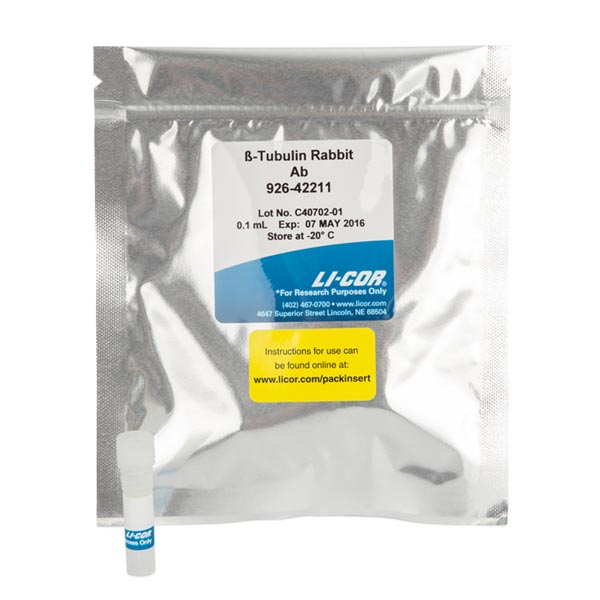
The β-Tubulin primary antibody can be used as an internal loading control (ILC) for normalization.
The expression of β-tubulin, or any housekeeping protein (HKP), should be validated to ensure that its expression does not change under experimental conditions.
Once validated, β-tubulin primary antibodies can be used for the detection of β-tubulin when performing two-color detection.
Detect β-Tubulin Rabbit Polyclonal Antibody with IRDye® Goat anti-Rabbit or IRDye Donkey anti-Rabbit secondary antibodies.
Other options for housekeeping protein normalization
β-微管蛋白一抗可用作内部上样对照 (ILC) 以进行标准化。
应验证 β-微管蛋白或任何管家蛋白 (HKP) 的表达,以确保其表达在实验条件下不会改变。
经验证后,β-微管蛋白一抗可在进行双色检测时用于检测 β-微管蛋白。
使用 IRDye® 山羊抗兔或 IRDye 驴抗兔二抗检测 β-微管蛋白兔多克隆抗体。
管家蛋白质标准化的其他选择
β-Tubulin antibody is supplied in 10 mM HEPES (pH 7.5), 150 mM NaCl, 100 µg/mL BSA, 50% glycerol, and <0.02% sodium azide.
Do not aliquot the antibody.
| Properties | β-Tubulin Rabbit Polyclonal Antibody (P/N 926-42211) |
|---|---|
| Species Cross-Reactivity | Human, mouse, rat, monkey, bovine |
| Target Molecular Weight | 55 kDa |
| Isotype | Rabbit IgG |
| Specificity/Sensitivity | Detects endogenous levels of total β-tubulin protein. Does not cross-react with recombinant α-tubulin. |
| Immunogen | A synthetic peptide (KLH-coupled) that corresponds to the sequence of total β-tubulin, and does not cross-react with recombinant α-tubulin |
| Tested Applications | Western blot (WB), Immunohistochemistry (IHC), Immunofluorescence (IF), Flow Cytometry (F)
蛋白质印迹 (WB)、免疫组织化学 (IHC)、免疫荧光 (IF)、流式细胞术 (F) |
