LI-COR IRDye® 700 p53 ,IRDye® 700 p53 共有寡核苷酸

Reagents > IRDye® 700 p53 Consensus Oligonucleotide,IRDye® 700 p53 共有寡核苷酸

IRDye® 700 p53 Consensus Oligonucleotide

5′ — TAC AGA ACA TGT CTA AGC ATG CTG GGG ACT — 3′

3′ — ATG TCT TGT ACA GAT TCG TAC GAC CCC TGA — 5′

Underlined nucleotides are the binding site. IRDye 700 oligonucleotides are supplied as 25 µL of 50 nM (or 50 fmol/µL) double-stranded DNA.

p53 is a cellular tumor antigen that functions as a tumor suppresor. For this reason, it is a common oligonucleotide used in EMSA to assess protein binding in cancer and related research.

带下划线的核苷酸是结合位点。 IRDye 700 寡核苷酸以 25 µL 的 50 nM(或 50 fmol/µL)双链 DNA 形式提供。

p53 是一种细胞肿瘤抗原,起肿瘤抑制作用。 因此,它是 EMSA 中常用的寡核苷酸,用于评估癌症和相关研究中的蛋白质结合。

You should establish conditions of the binding reaction for each protein-DNA pair. For IRDye 700 p53 oligonucleotide, the following binding reaction is a good starting point:

Reaction µL
10X Binding Buffer (100 mM Tris, 500 mM KCI, 10 mM DTT; pH 7.5) 2
Poly (dl•dC) 1 µg/µL in 10 mM Tris, 1 mM EDTA; pH 7.5 1
25 mM DTT/2.5% Tween® 20 2
100 mM McCl2 1
Water 12
IRDye 700 p53 1
HeLa 4 hour Serum Response nuclear extract (positive control) (5 µg/µL) 1
Total 20

After the addition of the DNA to the protein-buffer mix, reactions are incubated to allow protein binding to DNA. A typical incubation condition is 20-30 minutes at room temperature.

Since IRDye infrared dyes are somewhat sensitive to light, it is best to keep binding reactions in the dark during incubation periods (e.g., put the tubes into a drawer or simply cover the rack containing tubes with aluminum foil). After the incubation period, 10X Orange Loading Dye (P/N 927-10100) is added to the binding reaction for electrophoresis.

将 DNA 添加到蛋白质缓冲液混合物中后,孵育反应物以使蛋白质与 DNA 结合。 典型的孵育条件是室温下 20-30 分钟。

由于 IRDye 红外染料对光有些敏感,因此最好在孵育期间将结合反应保持在黑暗中(例如,将试管放入抽屉或简单地用铝箔盖住装有试管的架子)。 孵育期后,将 10X Orange Loading Dye (P/N 927-10100) 添加到结合反应中进行电泳。

LI-COR IRDye® 700,IRDye® 700 亚磷酰胺,LI-COR 4300 DNA

Reagents > IRDye® 700 Phosphoramidite,IRDye® 700 亚磷酰胺

IRDye® 700 Phosphoramidite

IRDye 700 Phosphoramidite has been optimized for the 700 nm channel of the LI-COR 4300 DNA Analysis System.

IRDye 700 亚磷酰胺针对 LI-COR 4300 DNA 分析系统的 700 nm 通道进行了优化。

IRDye Infrared Dyes Overview

Dye Structure

IRDye® 700 Phosphoramidite

Properties

  • Chemical Formula: C52H67N4O5PS
  • Molecular Weight: 891.16 g/mol
  • Exact Mass: 890.46

Absorption and Emission Spectra

Solvent Ext. Coeff. (M-1cm-1) Absorption Maxima Emission Maxima Stokes Shift
Methanol 170,000 680 nm 17 nm
Water 170,000 685 nm 705 nm 20 nm

Absorption and Emission Spectra in Water

IRDye® 700 Phosphoramidite

LI-COR P/N 927-10100,LI-COR IRDye® 700 Sp-1,IRDye® 700 Sp-1 共有寡核苷酸

Reagents > IRDye® 700 Sp-1 Consensus Oligonucleotide,IRDye® 700 Sp-1 共有寡核苷酸

IRDye® 700 Sp-1 Consensus Oligonucleotide

5′ — ATT CGA TCG GGG CGG GGC GAG C — 3′

3′ — TAA GCT AGC CCC GCC CCG CTC G — 5′

Underlined nucleotides are the binding site. IRDye 700 oligonucleotides are supplied as 25 µL of 50 nM (or 50 fmol/µL) double-stranded DNA.

Sp-1 is a transcription factor that regulates gene expression and controls a number of cellular processes including differentiation, proliferation, and apoptosis. For this reason, it is a common oligonucleotide used in EMSA to assess certain types protein binding.

带下划线的核苷酸是结合位点。 IRDye 700 寡核苷酸以 25 µL 的 50 nM(或 50 fmol/µL)双链 DNA 形式提供。

Sp-1 是一种转录因子,可调节基因表达并控制许多细胞过程,包括分化、增殖和凋亡。 出于这个原因,它是 EMSA 中用于评估某些类型的蛋白质结合的常见寡核苷酸。

You should establish conditions of the binding reaction for each protein-DNA pair. For IRDye 700 Sp-1 oligonucleotide, the following binding reaction is a good starting point:

Reaction µL
10X Binding Buffer (100 mM Tris, 500 mM KCI, 10 mM DTT; pH 7.5) 2
Poly (dl•dC) 1 µg/µL in 10 mM Tris, 1 mM EDTA; pH 7.5 1
25 mM DTT/2.5% Tween® 20 2
Water 13
IRDye 700 Sp-1 1
HeLa 4 hour Serum Response nuclear extract (positive control) (5 µg/µL) in Dilution Buffer (20 mM Hepes (pH 7.9), 100 mM KCI, 1 mM MgCl2, 20% glycerol, 0.5 mM PMSF, and 0.5 mM DTT) 1
Total 20

After the addition of the DNA to the protein-buffer mix, reactions are incubated to allow protein binding to DNA. A typical incubation condition is 20-30 minutes at room temperature.

Since IRDye infrared dyes are somewhat sensitive to light, it is best to keep binding reactions in the dark during incubation periods (e.g., put the tubes into a drawer or simply cover the rack containing tubes with aluminum foil). After the incubation period, 10X Orange Loading Dye (P/N 927-10100) is added to the binding reaction for electrophoresis.

将 DNA 添加到蛋白质缓冲液混合物中后,孵育反应物以使蛋白质与 DNA 结合。 典型的孵育条件是室温下 20-30 分钟。

由于 IRDye 红外染料对光有些敏感,因此最好在孵育期间将结合反应保持在黑暗中(例如,将试管放入抽屉或简单地用铝箔盖住装有试管的架子)。 孵育期后,将 10X Orange Loading Dye (P/N 927-10100) 添加到结合反应中进行电泳。

LI-COR P/N 927-10100,LI-COR IRDye® 700 STAT3,IRDye® 700 STAT3 共有寡核苷酸

Reagents > IRDye® 700 STAT3 Consensus Oligonucleotide,IRDye® 700 STAT3 共有寡核苷酸

IRDye® 700 STAT3 Consensus Oligonucleotide

5′ — GAT CCT TCT GGG AAT TCC TAG ATC — 3′

3′ — CTA GGA AGA CCC TTA AGG ATC TAG — 5′

Underlined nucleotides are the binding site. IRDye 700 oligonucleotides are supplied as 25 µL of 50 nM (or 50 fmol/µL) double-stranded DNA.

STAT3 is a transcription factor that is activated by phosphorylation of tyrosine 705 and in response to particular ligands. For this reason, it is a common oligonucleotide used in EMSA to assess certain types of protein binding.

You should establish conditions of the binding reaction for each protein-DNA pair. For IRDye 700 STAT3 oligonucleotide, the following binding reaction is a good starting point:

Reaction µL
10X Binding Buffer (100 mM Tris, 500 mM KCI, 10 mM DTT; pH 7.5) 2
Poly (dl•dC) 1 µg/µL in 10 mM Tris, 1 mM EDTA; pH 7.5 1
25 mM DTT/2.5% Tween® 20 2
1% NP-40 1
100 mM MgCl2 1
Water 11
IRDye 700 STAT3 1
U937 (IF Nα treated) nuclear extract (positive control) (5 µg/µL) 1
Total 20

After the addition of the DNA to the protein-buffer mix, reactions are incubated to allow protein binding to DNA. A typical incubation condition is 20-30 minutes at room temperature.

Since IRDye infrared dyes are somewhat sensitive to light, it is best to keep binding reactions in the dark during incubation periods (e.g., put the tubes into a drawer or simply cover the rack containing tubes with aluminum foil). After the incubation period, 10X Orange Loading Dye (P/N 927-10100) is added to the binding reaction for electrophoresis.

将 DNA 添加到蛋白质缓冲液混合物中后,孵育反应物以使蛋白质与 DNA 结合。 典型的孵育条件是室温下 20-30 分钟。

由于 IRDye 红外染料对光有些敏感,因此最好在孵育期间将结合反应保持在黑暗中(例如,将试管放入抽屉或简单地用铝箔盖住装有试管的架子)。 孵育期后,将 10X Orange Loading Dye (P/N 927-10100) 添加到结合反应中进行电泳。

LI-COR IRDye® 750,IRDye® 750 马来酰亚胺 红外染料750 nm

Reagents > IRDye® 750 Maleimide,IRDye® 750 马来酰亚胺

IRDye® 750 Maleimide

IRDye 750 Maleimide is an infrared dye with detection near 750 nm. Maleimide reactive group provides the functionality for labeling molecules that contain free sulfhydryl (–SH thiol) groups. This reactive group allows conjugation reactions to be performed at physiological pH.

IRDye 750 Maleimide 是一种红外染料,检测波长接近 750 nm。 马来酰亚胺反应基团提供了标记含有游离巯基 (-SH 硫醇) 基团的分子的功能。 该反应基团允许在生理 pH 下进行偶联反应。

This dye is not suitable for use with LI-COR Odyssey® Imaging Systems.

该染料不适用于 LI-COR Odyssey® 成像系统。

IRDye® 750 Maleimide

IRDye Infrared Dyes Overview

Dye Structure

IRDye® 750 Maleimide

Properties

  • Chemical Formula: C55H63N4Na3O15S4
  • Molecular Weight: 1217.33 g/mol
  • Exact Mass: 1216.29

Absorption and Emission Spectra

Solvent Ext. Coeff. (M-1cm-1) Absorption Maxima Emission Maxima
Methanol 330,000 766 nm 776 nm
1X PBS 260,000 756 nm 776 nm
PBS: Methanol 260,000

Absorption and Emission Spectra in 1X PBS

IRDye® 750 Maleimide

LI-COR IRDye® 750 NHS,IRDye® 750 NHS 酯 红外染料750 nm

Reagents > IRDye® 750 NHS Ester,IRDye® 750 NHS 酯

IRDye® 750 NHS Ester

IRDye 750 NHS Ester is an infrared dye with detection near 750 nm. The NHS ester reactive group provides the functionality for labeling primary and secondary amines, such as lysine residues in proteins.

IRDye 750 NHS Ester 是一种红外染料,检测波长接近 750 nm。 NHS 酯反应基团提供了标记伯胺和仲胺的功能,例如蛋白质中的赖氨酸残基。

This dye is not suitable for use with LI-COR Odyssey® Imaging Systems.

IRDye® 750 NHS Ester
NHS Ester reacts with a primary aliphatic amine, such as lysine.
IRDye® 750 NHS Ester
Dye label is attached through amide bond.

IRDye Infrared Dyes Overview

Dye Structure

IRDye® 750 NHS Ester

Properties

  • Chemical Formula: C53H60N3Na3O16S4
  • Molecular Weight: 1192.28 g/mol
  • Exact Mass: 1191.25

Absorption and Emission Spectra

Solvent Ext. Coeff. (M-1cm-1) Absorption Maxima Emission Maxima
Methanol 330,000 766 nm 776 nm
1X PBS 260,000 756 nm 776 nm
PBS: Methanol 260,000

Absorption and Emission Spectra in Methanol

IRDye® 750 NHS Ester

LI-COR 4300 DNA,LI-COR IRDye® 800,IRDye® 800 亚磷酰胺 800 nm

Reagents > IRDye® 800 Phosphoramidite,IRDye® 800 亚磷酰胺

IRDye® 800 Phosphoramidite

IRDye 800 Phosphoramidite has been optimized for the 800 nm channel of the LI-COR 4300 DNA Analysis System.

IRDye 800 亚磷酰胺针对 LI-COR 4300 DNA 分析系统的 800 nm 通道进行了优化。

IRDye Infrared Dyes Overview

Dye Structure

IRDye® 800 Phosphoramidite

Properties

  • Chemical Formula: C59H75N4O6PS
  • Molecular Weight: 999.30 g/mol
  • Exact Mass: 998.51

Absorption and Emission Spectra in Water

IRDye® 800 Phosphoramidite

LI-COR IRDye® 800CW 2-DG 光学探头,IRDye 800CW 2-脱氧葡萄糖

Reagents > IRDye® 800CW 2-DG Optical Probe,IRDye® 800CW 2-DG 光学探头

IRDye® 800CW 2-DG Optical Probe

IRDye 800CW 2-DG (2-deoxyglucose) is a fluorescent optical imaging agent that has been shown to be reactive with implanted tumors derived from many cell lines including A431, SW620, 3T3-L1, and PC3LMN4. This optical imaging agent has been used for numerous applications, including research pertaining to tumor biology, tumor metastases, diabetes, and arthritis.

Cancer cells are often characterized by a high metabolic rate exemplified by an elevated rate of glycolysis. This observation forms the basis for positron emission tomography (PET) using glucose analogues such as 18F-2-deoxy-D-glucose (18FDG) to visualize primary tumors and their metastasis. Radiolabeled 2-deoxy-D-glucose (2-DG) is generally impractical for use in small animal studies of cancer biology. Fluorophore-labeled variants of 2-deoxy-D-glucose have been used with varying degrees of success.1, 2, 3

The optical agent exhibits the expected dose response with increasing concentrations of the agent in both cell based and animal experiments. Uptake of the agent can be effectively blocked with either unlabeled 2-DG or glucose, confirming its specificity.

Confocal microscopy demonstrated conclusively that IRDye 800CW 2-DG is taken up by the cell and localized in the cytoplasm. While the exact mechanism of IRDye 800CW 2-DG uptake is not known, there is strong evidence for the involvement of the GLUT transporter system.

IRDye 800CW 2-DG(2-脱氧葡萄糖)是一种荧光光学成像剂,已被证明与来自许多细胞系(包括 A431、SW620、3T3-L1 和 PC3LMN4)的植入肿瘤发生反应。这种光学显像剂已用于许多应用,包括与肿瘤生物学、肿瘤转移、糖尿病和关节炎有关的研究。

癌细胞通常以高代谢率为特征,例如糖酵解率升高。该观察结果构成了使用葡萄糖类似物(例如 18F-2-脱氧-D-葡萄糖 (18FDG))可视化原发性肿瘤及其转移的正电子发射断层扫描 (PET) 的基础。放射性标记的 2-脱氧-D-葡萄糖 (2-DG) 通常不适用于癌症生物学的小动物研究。使用荧光团标记的 2-脱氧-D-葡萄糖变体已取得不同程度的成功。 1, 2, 3

在基于细胞的实验和动物实验中,随着试剂浓度的增加,光学试剂表现出预期的剂量反应。未标记的 2-DG 或葡萄糖可以有效地阻断该试剂的摄取,从而证实了其特异性。

共聚焦显微镜最终证明 IRDye 800CW 2-DG 被细胞吸收并定位于细胞质中。虽然 IRDye 800CW 2-DG 摄取的确切机制尚不清楚,但有强有力的证据表明 GLUT 转运系统参与其中。

Learn about other LI-COR optical probes.

Applications

  • Microscopy
  • Optical Probe Development
  • Target Specificity
  • Tissue Section Imaging
  • 显微镜
    光学探头开发
    目标特异性
    组织切片成像

Absorbance and Emission Spectra

IRDye® 800CW 2-DG Optical Probe

Data Example

IRDye® 800CW 2-DG Optical Probe
Two examples in which IRDye 800CW 2-DG Optical Probe (10 nmol) was used to detect subcutaneous A431 tumors. Probe signal is displayed in pseudo color and is merged with a grey scale white light image of the mouse.
使用 IRDye 800CW 2-DG 光学探头 (10 nmol) 检测皮下 A431 肿瘤的两个示例。 探针信号以伪彩色显示,并与鼠标的灰度白光图像合并。

References

  1. Lloyd, P.G., Hardin, C.D., and Sturek, M. (1999). Physiol Res, 48. 401-10.
  2. O’Neil, R.G., Wu, L., and Mullani, N. (2005) Mol Imaging Biol, 7. 388-92.
  3. Cheng, Z., Levi, J., Xiong, Z., Gheysens, O., Keren, S., Chen, X., and Gambhir, S.S. (2006) Bioconjug Chem, 17. 662-9.
  4. Kovar, J. L., Volcheck, W. M., Sevick-muraca, E., Simpson, M. A., & Olive, D. M. (2009). Anal Biochem, 384(2). 254-262. doi: 10.1016/j.ab.2008.09.050.

LI-COR IRDye 800CW ,IRDye® 800CW 炔烃红外染料

Reagents > IRDye® 800CW Alkyne Infrared Dye,IRDye® 800CW 炔烃红外染料

IRDye® 800CW Alkyne Infrared Dye

IRDye 800CW Alkyne provides the functionality for preferential labeling of molecules containing azide groups (–N3) with IRDye 800CW.

IRDye 800CW Alkyne 提供了使用 IRDye 800CW 优先标记含有叠氮基 (–N3) 的分子的功能。

Learn more about Near-Infrared Fluorescent Click Chemistry Reagents.

Dye Structure

IRDye® 800CW Alkyne Infrared Dye

Properties

  • Chemical Formula: C49H54N3Na3O14S4
  • Molecular Weight: 1106.19 g/mol
  • Exact Mass: 1105.22
  • Correction Factor at 260 – 280 nm: 3%

Absorption and Emission Spectra

Solvent Absorption Maxima Emission Maxima
Methanol 778 nm 794 nm
1X PBS 774 nm 789 nm
1:1 Methanol to 1X PBS 778 nm

Absorbance and Emission Spectra in 1X PBS

IRDye® 800CW Alkyne Infrared Dye

Molar Absorptivity

Solvent Molar Absorptivity (L mol-1 cm-1)
Methanol 300,000
1X PBS 240,000
1:1 Methanol to 1X PBS 270,000

LI-COR IRDye® 800CW,IRDye® 800CW 叠氮化物红外染料

Reagents > IRDye® 800CW Azide Infrared Dye,IRDye® 800CW 叠氮化物红外染料

IRDye® 800CW Azide Infrared Dye

IRDye 800CW Azide provides the functionality for preferential labeling of molecules that contain either the alkyne or dibenzocyclooctyne (DBCO) group.

IRDye 800CW 叠氮化物提供优先标记含有炔烃或二苯并环辛炔 (DBCO) 基团的分子的功能。

Learn more about Near-Infrared Fluorescent Click Chemistry Reagents.

Dye Structure

IRDye® 800CW Azide Infrared Dye

Properties

  • Chemical Formula: C54H67N6Na3O17S4
  • Molecular Weight: 1269.36 g/mol
  • Exact Mass: 1268.31
  • Correction factor at 260 – 280 nm: 3%

Absorption and Emission Spectra

Solvent Absorption Maxima Emission Maxima
Methanol 778 nm 794 nm
1X PBS 774 nm 789 nm
1:1 Methanol to 1X PBS 778 nm

Absorbance and Emission Spectra in 1X PBS

IRDye® 800CW Azide Infrared Dye

Molar Absorptivity

Solvent Molar Absorptivity (L mol-1 cm-1)
Methanol 300,000
1X PBS 240,000
1:1 Methanol to 1X PBS 270,000

Example Data

IRDye® 800CW Azide Infrared Dye
Palmitoylation of Lck kinase analyzed with a Click Chemistry IRDye reagent.​​ Lck was immunoprecipitated from metabolically-labeled Jurkat cell lysates. Palmitoylation was detected with CuAAC click chemistry and IRDye 800CW Azide Infrared Dye​ (green signals; P/N 929-60000). Total Lck was detected with anti-Lck D88 antibody and IRDye 680RD Donkey anti-Rabbit IgG​ (red; P/N 926-68073). Yellow indicates overlap of signals. The mobility shift caused by palmitoylation is observed in lanes 3-5 and 7-9 (green bands migrating more slowly than red bands). Data and image supplied by a LI-COR customer.
使用 Click Chemistry IRDye 试剂分析 Lck 激酶的棕榈酰化。Lck 从代谢标记的 Jurkat 细胞裂解物中进行免疫沉淀。 使用 CuAAC 点击化学和 IRDye 800CW 叠氮化物红外染料(绿色信号;P/N 929-60000)检测棕榈酰化。 使用抗 Lck D88 抗体和 IRDye 680RD 驴抗兔 IgG(红色;P/N 926-68073)检测总 Lck。 黄色表示信号重叠。 在泳道 3-5 和 7-9 中观察到由棕榈酰化引起的迁移率变化(绿色条带迁移速度比红色条带慢)。 LI-COR 客户提供的数据和图像。

LI-COR IRDye® 800CW BoneTag™光学探头,IRDye 800CW 近红外染料

Reagents > IRDye® 800CW BoneTag™ Optical Probe,IRDye® 800CW BoneTag™光学探头

IRDye® 800CW BoneTag™  Optical Probe

IRDye 800CW BoneTag Optical Probe is a calcium-chelating compound conjugated to IRDye 800CW near-infrared dye. Using IRDye NIR dyes extends fluorescence signal detection to the near-infrared fluorescent region of the spectrum without affecting the compound’s ability to function as a marker of the mineralization process. NIR fluorescence detection improves depth of penetration due to low tissue autofluorescence, translating to low background interference.

Calcium-chelating compounds have been used effectively for the detection of bone mineralization, growth, and morphological changes, including tetracycline derivatives, xylenol orange, alizarin, calcein, and fluorescein. NIR-labeled pamidronate has also been successfully used for detection of arterial calcification in non-invasive optical imaging1.

The ability to visualize bone anatomy and structures for an extended period facilitates use of the IRDye BoneTag in conjunction with a second optical agent specific for a primary target (i.e., tumor tissue). In addition, IRDye 800CW BoneTag may prove effective for studies dealing with bone metastasis and bone remodeling studies.

IRDye 800CW BoneTag 光学探针是一种与 IRDye 800CW 近红外染料结合的钙螯合化合物。使用 IRDye NIR 染料将荧光信号检测扩展到光谱的近红外荧光区域,而不会影响化合物作为矿化过程标记的能力。 NIR 荧光检测由于组织自发荧光低而提高了穿透深度,从而转化为低背景干扰。

钙螯合化合物已被有效地用于检测骨矿化、生长和形态变化,包括四环素衍生物、二甲酚橙、茜素、钙黄绿素和荧光素。 NIR 标记的帕米膦酸盐也已成功用于检测无创光学成像中的动脉钙化1。

长时间可视化骨骼解剖和结构的能力有助于将 IRDye BoneTag 与特定于主要目标(即肿瘤组织)的第二种光学试剂结合使用。此外,IRDye 800CW BoneTag 可能证明对处理骨转移和骨重塑研究的研究有效。

Learn about other LI-COR optical probes.

Applications

  • Microscopy
  • Bone Imaging

Absorbance and Emission Spectra

IRDye® 800CW BoneTag™  Optical Probe

Data Example

IRDye® 800CW BoneTag™  Optical Probe
Bone-targeted binding was examined using MC3T3 (osteoblast) cells in a cell-based assay. MC3T3 cells exhibited a dose dependent increase in signal when incubated with IRDye 800CW BoneTag. Incubation of labeled BoneTag with A431 cells (epidermoid carcinoma), a non-osteoblast cell line, showed low non-specific binding.
在基于细胞的测定中使用 MC3T3(成骨细胞)细胞检查骨靶向结合。 当与 IRDye 800CW BoneTag 一起孵育时,MC3T3 细胞表现出剂量依赖性的信号增加。 标记的 BoneTag 与非成骨细胞系 A431 细胞(表皮样癌)的孵育显示出低非特异性结合。

References

  1. Zaheer A et al. (2001) In vivo near-infrared fluorescence imaging of osteoblastic activity. Nat Biotechnol, 19. 1148-54.
  2. Kovar, J. L., Xu, X., Draney, D., Cupp, A., Simpson, M. A., & Michael Olive, D. (2011). Anal Biochem, 416(2), 167–173. doi: 10.1016/j.ab.2011.05.011.

LI-COR IRDye® 800CW 羧酸盐,IRDye 800CW

Reagents > IRDye® 800CW Carboxylate,IRDye® 800CW 羧酸盐

IRDye® 800CW Carboxylate

Assays that use IRDye 800CW conjugates (such as in vivo imaging and cell binding assays) may require a “dye-only” control for potential effects or retention of the dye.

Carboxylate (non-reactive) form of IRDye 800CW is an ideal control.

使用 IRDye 800CW 偶联物的检测(例如体内成像和细胞结合检测)可能需要“仅染料”对照来控制染料的潜在影响或保留。

IRDye 800CW 的羧酸盐(非反应性)形式是理想的对照。

Note: The carboxylate dye has no reactive group and cannot be used for labeling.

IRDye® 800CW Carboxylate
Clearance kinetics of IRDye 800CW carboxylate. A single SCID mouse was injected with 1 nmol of non-reactive IRDye 800CW dye, and clearance monitored over time as indicated. Pseudocolor fluorescence is superimposed on a white light image. IRDye 800CW carboxylate dispersed rapidly and was completely cleared after 48 h. Originally published in Kovar, J., et al. (2007) Anal Biochem 367:1-12.
IRDye 800CW 羧酸盐的清除动力学。 给一只 SCID 小鼠注射 1 nmol 的非反应性 IRDye 800CW 染料,并如图所示随时间监测清除率。 伪彩色荧光叠加在白光图像上。 IRDye 800CW 羧酸盐迅速分散,48 小时后完全清除。 最初发表于 Kovar, J. 等人。 (2007) 肛门生物化学 367:1-12。

How is IRDye 800CW Carboxylate Used?

In vitro Cell-based Assays

As a control for cell-based assays that monitor binding of a dye-labeled agent.

  • Validation of optical agents for in vivo administration
  • Evaluation of binding specificity

In vivo Imaging

For evaluation of behavior and clearance of the dye itself.

  • Timing of dye clearance from the animal’s body
  • Retention of dye in certain organs or sites (e.g., liver or kidneys)

Labeling Reaction Reference

As a standard to determine the amount of unreacted (“free”) dye after IRDye 800CW conjugation and purification.

Residual unreacted dye may cause:

  • Artificially high values when dye/protein (D/P) ratio is calculated
  • Increased background fluorescence in biological assays

Dye Structure

IRDye® 800CW Carboxylate

Properties

  • Chemical Formula: C46H50N2Na4O15S4
  • Molecular Weight: 1091.10 g/mol
  • Exact Mass: 1090.17

LI-COR IRDye® 800CW DBCO 红外染料,IRDye® 800CW DBCO

Reagents > IRDye® 800CW DBCO Infrared Dye,IRDye® 800CW DBCO 红外染料

IRDye® 800CW DBCO Infrared Dye

IRDye 800CW DBCO provides the functionality for preferential labeling of molecules that contain azide groups (–N3) with IRDye 800CW.

IRDye 800CW DBCO 提供了使用 IRDye 800CW 优先标记含有叠氮基 (–N3) 的分子的功能。

Learn more about Near-Infrared Fluorescent Click Chemistry Reagents.

Dye Structure

IRDye® 800CW DBCO Infrared Dye

Properties

  • Chemical Formula: C64H65N4Na3O15S4
  • Molecular Weight: 1327.45 g/mol
  • Exact Mass: 1326.30
  • Correction Factor at 260 – 280 nm: 3%

Absorption and Emission Spectra

Solvent Absorption Maxima Emission Maxima
Methanol 778 nm 794 nm
1X PBS 774 nm 789 nm
1:1 Methanol to 1X PBS 778 nm

Absorbance and Emission Spectra in 1X PBS

IRDye® 800CW DBCO Infrared Dye

Molar Absorptivity

Solvent Molar Absorptivity (L mol-1 cm-1)
Methanol 300,000
1X PBS 240,000
1:1 Methanol to 1X PBS 270,000

LI-COR IRDye 800CW 二抗,LI-COR IRDye® 800CW 驴抗鸡二抗

Reagents > IRDye® 800CW Donkey anti-Chicken Secondary Antibody,IRDye® 800CW 驴抗鸡二抗

IRDye® 800CW Donkey anti-Chicken Secondary Antibody

Immunogen

Chicken IgY, whole molecule. IgY is the original designation for the IgG-like protein found in both serum and egg yolk.

Purity and Specificity

The antibody was isolated from antisera by immunoaffinity chromatography using antigens coupled to agarose beads. Based on immunoelectrophoresis and/or ELISA, this antibody reacts with whole molecule chicken IgY, and with the light chains of other chicken immunoglobulins. No reactivity was detected against non-immunoglobulin serum proteins. This antibody was tested by ELISA and/or solid-phase adsorbed to ensure minimal cross-reactivity with bovine, goat, guinea pig, Syrian hamster, horse, human, mouse, rabbit, rat, and sheep serum proteins, but may cross-react with immunoglobulins from other species. The conjugate has been specifically tested and qualified for Western blot applications.

使用与琼脂糖珠偶联的抗原,通过免疫亲和层析从抗血清中分离抗体。 基于免疫电泳和/或 ELISA,该抗体与全分子鸡 IgY 以及其他鸡免疫球蛋白的轻链反应。 未检测到针对非免疫球蛋白血清蛋白的反应性。 该抗体通过 ELISA 和/或固相吸附测试,以确保与牛、山羊、豚鼠、叙利亚仓鼠、马、人、小鼠、兔、大鼠和绵羊血清蛋白的交叉反应最小,但可能发生交叉反应 与来自其他物种的免疫球蛋白。 该偶联物经过专门测试,可用于蛋白质印迹应用。

Applications

Highly recommended for:

  • Western Blot
  • In-Cell Western™ Assay
  • On-Cell Western Assay
  • Protein Array
  • Immunohistochemistry
  • Small Animal Imaging
  • Microscopy
  • 2D Gel Detection
  • Tissue Section Imaging
  • Virus Titration Assay
  • 蛋白质印迹
    In-Cell Western™ 检测
    On-Cell Western 检测
    蛋白质阵列
    免疫组织化学
    小动物成像
    显微镜
    2D 凝胶检测
    组织切片成像
    病毒滴定法

Formulation

IRDye 800CW secondary antibodies are supplied as purified immunoglobulin conjugates, lyophilized in phosphate-buffered saline, pH 7.4. Protect from light. Store at 4 °C prior to reconstitution.

Each vial contains 10 mg/mL BSA (free of IgG and protease) as a stabilizer and 0.01% sodium azide as a preservative, after reconstitution. Concentration is 1.0 mg/mL when reconstituted as directed. Refer to the pack insert for details on reconstitution.

IRDye 800CW 二抗以纯化的免疫球蛋白偶联物形式提供,在 pH 7.4 的磷酸盐缓冲盐水中冻干。 避光。 在重组前储存在 4°C。

复溶后,每瓶含有 10 mg/mL BSA(不含 IgG 和蛋白酶)作为稳定剂和 0.01% 叠氮化钠作为防腐剂。 按照指示重新配制时,浓度为 1.0 mg/mL。 有关重组的详细信息,请参阅包装插页。

Recommended Dilutions

Application Recommended Suggested Range
Odyssey Western blot detection 1:15,000 1:5,000 – 1:25,000
Other User optimized

Optimum dilutions will vary and should be determined empirically.

RRID

  • P/N 925-32218: RRID AB_2814922
  • P/N 926-32218: RRID AB_1850023

Example Data

IRDye® 800CW Donkey anti-Chicken Secondary Antibody
Detection of actin with IRDye 800CW Donkey anti-chicken. Odyssey® One-Color Molecular Weight Marker (P/N 928-40000) and C32 lysate, 3 µg – 0.01 µg (Santa Cruz P/N sc-2205) were resolved on a 10% Bis Tris gel and transferred to Odyssey Nitrocellulose membrane (P/N 926-31092). Membrane was blocked with Odyssey Blocking Buffer (P/N 927-40000) followed by detection with chicken anti-actin (GenWay P/N 15-288-23014) and IRDye 800CW Donkey anti-Chicken (P/N 926-32218).
用 IRDye 800CW 驴抗鸡检测肌动蛋白。 Odyssey® One-Color Molecular Weight Marker (P/N 928-40000) 和 C32 裂解物,3 µg – 0.01 µg (Santa Cruz P/N sc-2205) 在 10% Bis Tris 凝胶上分离并转移到 Odyssey 硝酸纤维素膜上 (部件号 926-31092)。 用 Odyssey 封闭缓冲液 (P/N 927-40000) 封闭膜,然后用鸡抗肌动蛋白 (GenWay P/N 15-288-23014) 和 IRDye 800CW Donkey anti-Chicken (P/N 926-32218) 进行检测。

LI-COR IRDye 800CW 二抗,LI-COR IRDye® 800CW 驴抗山羊 IgG 二抗,

Reagents > IRDye® 800CW Donkey anti-Goat IgG Secondary Antibody, IRDye® 800CW 驴抗山羊 IgG 二抗

IRDye® 800CW Donkey anti-Goat IgG Secondary Antibody

Immunogen

Goat IgG, whole molecule

Purity and Specificity

The antibody was isolated from antisera by immunoaffinity chromatography using antigens coupled to agarose beads. Based on electrophoresis, this antibody reacts with the heavy and light chains of goat IgG, and with the light chains common to most goat immunoglobulins. This antibody also reacts with sheep IgG. No antibody was detected against non-immunoglobulin serum proteins. This antibody has been tested by ELISA and/or solid-phase adsorbed to ensure minimal cross-reactivity with chicken. guinea pig, Syrian hamster, horse, human, mouse, rat, and rabbit serum proteins, but may cross-react with immunoglobulins from other species. The conjugate has been specifically tested and qualified for Western blot applications.

使用与琼脂糖珠偶联的抗原,通过免疫亲和层析从抗血清中分离抗体。 基于电泳,该抗体与山羊 IgG 的重链和轻链以及大多数山羊免疫球蛋白共有的轻链发生反应。 该抗体还与绵羊 IgG 发生反应。 未检测到针对非免疫球蛋白血清蛋白的抗体。 该抗体已通过 ELISA 和/或固相吸附测试,以确保与鸡的交叉反应最小。 豚鼠、叙利亚仓鼠、马、人、小鼠、大鼠和兔血清蛋白,但可能与其他物种的免疫球蛋白发生交叉反应。 该偶联物经过专门测试,可用于蛋白质印迹应用。

Applications

Highly recommended for:

  • Western Blot
  • In-Cell Western™ Assay
  • On-Cell Western Assay
  • Protein Array
  • Immunohistochemistry
  • Small Animal Imaging
  • Microscopy
  • 2D Gel Detection
  • Tissue Section Imaging
  • Virus Titration Assay

Note: Do not use this secondary antibody in combination with any secondary antibody whose host is goat (for example, IRDye 680RD Goat anti-Rabbit) for two-color Western blot detection.

Formulation

IRDye 800CW secondary antibodies are supplied as purified immunoglobulin conjugates, lyophilized in phosphate-buffered saline, pH 7.4. Protect from light. Store at 4 °C prior to reconstitution.

Each vial contains 10 mg/mL BSA (free of IgG and protease) as a stabilizer and 0.01% sodium azide as a preservative, after reconstitution. Concentration is 1.0 mg/mL when reconstituted as directed. Refer to the pack insert for details on reconstitution.

IRDye 800CW 二抗以纯化的免疫球蛋白偶联物形式提供,在 pH 7.4 的磷酸盐缓冲盐水中冻干。 避光。 在重组前储存在 4°C。

复溶后,每瓶含有 10 mg/mL BSA(不含 IgG 和蛋白酶)作为稳定剂和 0.01% 叠氮化钠作为防腐剂。 按照指示重新配制时,浓度为 1.0 mg/mL。 有关重组的详细信息,请参阅包装插页。

Recommended Dilutions

Application Recommended Suggested Range
Odyssey® Western blot detection 1:15,000 1:5,000 – 1:25,000
Other User optimized

Optimum dilutions will vary and should be determined empirically.

RRID

  • P/N 925-32214: RRID AB_2687553
  • P/N 926-32214: RRID AB_621846

LI-COR IRDye® 800CW 驴抗豚鼠 IgG 二抗,In-Cell Western™ 检测

Reagents > IRDye® 800CW Donkey anti-Guinea Pig IgG Secondary Antibody,IRDye® 800CW 驴抗豚鼠 IgG 二抗

IRDye® 800CW Donkey anti-Guinea Pig IgG Secondary Antibody

Immunogen

Guinea Pig IgG, whole molecule

Purity and Specificity

Isolation of specific antibodies was accomplished by affinity chromatography using antigens immobilized on agarose beads. Based on immunoelectrophoresis and/or ELISA, this antibody reacts with whole molecule guinea pig IgG as well as the light chains common to other guinea pig immunoglobulins. No reactivity was detected against non-immunoglobulin serum proteins. This antibody has been tested by ELISA and/or solid-phase adsorbed to ensure minimal cross-reactivity with bovine, chicken, goat, Syrian hamster, horse, human, mouse, rabbit, rat, and sheep serum proteins. The conjugate has been specifically tested and qualified for Western blot and In-Cell Western™ assay applications.

使用固定在琼脂糖珠上的抗原通过亲和层析实现特异性抗体的分离。 基于免疫电泳和/或 ELISA,该抗体与全分子豚鼠 IgG 以及其他豚鼠免疫球蛋白共有的轻链反应。 未检测到针对非免疫球蛋白血清蛋白的反应性。 该抗体已通过 ELISA 和/或固相吸附测试,以确保与牛、鸡、山羊、叙利亚仓鼠、马、人、小鼠、兔、大鼠和绵羊血清蛋白的交叉反应最小。 该偶联物经过专门测试,可用于蛋白质印迹和 In-Cell Western™ 检测应用。

Applications

Highly recommended for:

  • Western Blot
  • In-Cell Western Assay
  • On-Cell Western Assay
  • Protein Array
  • Immunohistochemistry
  • Small Animal Imaging
  • Microscopy
  • 2D Gel Detection
  • Tissue Section Imaging
  • Virus Titration Assay
  • 蛋白质印迹
    细胞内西方检测
    On-Cell Western 检测
    蛋白质阵列
    免疫组织化学
    小动物成像
    显微镜
    2D 凝胶检测
    组织切片成像
    病毒滴定法

Formulation

IRDye 800CW secondary antibodies are supplied as purified immunoglobulin conjugates, lyophilized in phosphate-buffered saline, pH 7.4. Protect from light. Store at 4 °C prior to reconstitution.

Each vial contains 10 mg/mL BSA (free of IgG and protease) as a stabilizer and 0.01% sodium azide as a preservative, after reconstitution. Concentration is 1.0 mg/mL when reconstituted as directed. Refer to the pack insert for details on reconstitution.

IRDye 800CW 二抗以纯化的免疫球蛋白偶联物形式提供,在 pH 7.4 的磷酸盐缓冲盐水中冻干。 避光。 在重组前储存在 4°C。

复溶后,每瓶含有 10 mg/mL BSA(不含 IgG 和蛋白酶)作为稳定剂和 0.01% 叠氮化钠作为防腐剂。 按照指示重新配制时,浓度为 1.0 mg/mL。 有关重组的详细信息,请参阅包装插页。

Recommended Dilutions

Application Recommended Suggested Range
Odyssey Western blot detection 1:15,000 1:5,000 – 1:25,000
In-Cell Western assay detection 1:800 1:200 – 1:1,200
Other User optimized

Optimum dilutions will vary and should be determined empirically.

RRID

  • P/N 925-32411: RRID AB_2814905
  • P/N 926-32411: RRID AB_1850024

Example Data

IRDye® 800CW Donkey anti-Guinea Pig IgG Secondary Antibody
Detection of guinea pig IgG with IRDye 800CW Donkey anti-Guinea Pig. Serial dilutions of Guinea Pig IgG were spiked into C32 lysate (Santa Cruz P/N sc-2205). Samples were resolved on a 10% Bis-Tris gel and transferred to Odyssey® Nitrocellulose membrane (P/N 926-31092). The membrane was blocked with Odyssey Blocking Buffer (P/N 927-40000) followed by detection with IRDye 800CW Donkey anti-Guinea Pig (P/N 926-32411).

LI-COR IRDye 800CW 二抗,LI-COR IRDye® 800CW 驴抗小鼠 IgG 二抗

Reagents > IRDye® 800CW Donkey anti-Mouse IgG Secondary Antibody,IRDye® 800CW 驴抗小鼠 IgG 二抗

IRDye® 800CW Donkey anti-Mouse IgG Secondary Antibody

Immunogen

Mouse IgG

Purity and Specificity

The antibody was isolated by affinity chromatography using antigens coupled to agarose beads. Based on ELISA, this antibody reacts with the heavy and light chains of mouse IgG, and with the light chains of mouse IgM and IgA. This antibody was tested by ELISA and/or solid-phase adsorbed to ensure minimal cross-reactivity with bovine, chicken, goat, guinea pig, horse, human, rabbit, and sheep serum proteins, but may cross-react with immunoglobulins from other species. The conjugate has been specifically tested and qualified for Western blot and In-Cell Western™ assay applications.

使用与琼脂糖珠偶联的抗原通过亲和层析分离抗体。 基于 ELISA,该抗体与小鼠 IgG 的重链和轻链以及小鼠 IgM 和 IgA 的轻链反应。 该抗体通过 ELISA 和/或固相吸附测试,以确保与牛、鸡、山羊、豚鼠、马、人、兔和绵羊血清蛋白的交叉反应最小,但可能与来自其他物种的免疫球蛋白交叉反应 . 该偶联物经过专门测试,可用于蛋白质印迹和 In-Cell Western™ 检测应用。

Applications

Highly Recommended for:

  • Western Blot
  • In-Cell Western Assay
  • On-Cell Western Assay
  • Protein Array
  • Immunohistochemistry
  • Small Animal Imaging
  • Microscopy
  • 2D Gel Detection
  • Tissue Section Imaging
  • Virus Titration Assay
  • 蛋白质印迹
    细胞内西方检测
    On-Cell Western 检测
    蛋白质阵列
    免疫组织化学
    小动物成像
    显微镜
    2D 凝胶检测
    组织切片成像
    病毒滴定法

Formulation

IRDye 800CW secondary antibodies are supplied as purified immunoglobulin conjugates, lyophilized in phosphate-buffered saline, pH 7.4. Protect from light. Store at 4 °C prior to reconstitution.

Each vial contains 10 mg/mL BSA (free of IgG and protease) as a stabilizer and 0.01% sodium azide as a preservative, after reconstitution. Concentration is 1.0 mg/mL when reconstituted as directed. Refer to the pack insert for details on reconstitution.

IRDye 800CW 二抗以纯化的免疫球蛋白偶联物形式提供,在 pH 7.4 的磷酸盐缓冲盐水中冻干。 避光。 在重组前储存在 4°C。

复溶后,每瓶含有 10 mg/mL BSA(不含 IgG 和蛋白酶)作为稳定剂和 0.01% 叠氮化钠作为防腐剂。 按照指示重新配制时,浓度为 1.0 mg/mL。 有关重组的详细信息,请参阅包装插页。

Recommended Dilutions

Application Recommended Suggested Range
Odyssey Western blot detection 1:15,000 1:5,000 – 1:25,000
In-Cell Western assay detection 1:800 1:200 – 1:800
Other User optimized

Optimum dilutions will vary and should be determined empirically.

RRID

  • P/N 925-32212: RRID AB_2716622
  • P/N 926-32212: RRID AB_621847

Example Data

IRDye® 800CW Donkey anti-Mouse IgG Secondary Antibody
Western blot analysis of PTEN expression in mouse PTEN transfected 293T whole cell lysate (lane 2) and non-transfected 293T lysate (lane 3). Both lysates were loaded with 2 µg total protein per lane. Odyssey® One-Color Molecular Weight Marker (P/N 928-40000) was loaded in lane 1. The blot was probed with mouse ant-PTEN (IgG2b) (Santa Cruz sc-133197) and chicken anti-GAPDH (ProSci XW-7214) followed by detection with IRDye 800CW Donkey anti-Mouse (P/N 926-32212) and IRDye 680LT Donkey anti-Chicken (P/N 926-68028).
小鼠 PTEN 转染的 293T 全细胞裂解物(泳道 2)和未转染的 293T 裂解物(泳道 3)中 PTEN 表达的蛋白质印迹分析。 两种裂解物每条泳道均加载 2 µg 总蛋白。 将 Odyssey® One-Color Molecular Weight Marker (P/N 928-40000) 加载到泳道 1。用小鼠 ant-PTEN (IgG2b) (Santa Cruz sc-133197) 和鸡抗 GAPDH (ProSci XW- 7214),然后使用 IRDye 800CW 驴防鼠 (P/N 926-32212) 和 IRDye 680LT 驴防鸡 (P/N 926-68028) 进行检测。

LI-COR IRDye® 800CW 驴抗兔 IgG 二抗,LI-COR IRDye 800CW 二抗

Reagents > IRDye® 800CW Donkey anti-Rabbit IgG Secondary Antibody,IRDye® 800CW 驴抗兔 IgG 二抗

IRDye® 800CW Donkey anti-Rabbit IgG Secondary Antibody

Immunogen

Rabbit IgG

Purity and Specificity

The antibody was isolated by affinity chromatography using antigens coupled to agarose beads. Based on ELISA, this antibody reacts with the heavy and light chains of rabbit IgG, and with the light chains common to most rabbit immunoglobulins. This antibody was tested by ELISA and/or solid-phase adsorbed to ensure minimal cross-reactivity with bovine, chicken, goat, guinea pig, hamster, horse, human, mouse, rat, and sheep serum proteins, but may cross-react with immunoglobulins from other species. The conjugate has been specifically tested and qualified for Western blot and In-Cell Western™ assay applications.

使用与琼脂糖珠偶联的抗原通过亲和层析分离抗体。 基于 ELISA,该抗体与兔 IgG 的重链和轻链以及大多数兔免疫球蛋白常见的轻链发生反应。 该抗体通过 ELISA 和/或固相吸附测试,以确保与牛、鸡、山羊、豚鼠、仓鼠、马、人、小鼠、大鼠和绵羊血清蛋白的交叉反应最小,但可能与 来自其他物种的免疫球蛋白。 该偶联物经过专门测试,可用于蛋白质印迹和 In-Cell Western™ 检测应用。

Applications

Highly recommended for:

  • Western Blot
  • In-Cell Western Assay
  • On-Cell Western Assay
  • FRET-Based Assays
  • Protein Array
  • Immunohistochemistry
  • Small Animal Imaging
  • Microscopy
  • 2D Gel Detection
  • Tissue Section Imaging
  • Virus Titration Assay
  • 蛋白质印迹
    细胞内西方检测
    On-Cell Western 检测
    基于 FRET 的检测
    蛋白质阵列
    免疫组织化学
    小动物成像
    显微镜
    2D 凝胶检测
    组织切片成像
    病毒滴定法

Formulation

IRDye 800CW secondary antibodies are supplied as purified immunoglobulin conjugates, lyophilized in phosphate-buffered saline, pH 7.4. Protect from light. Store at 4 °C prior to reconstitution.

Each vial contains 10 mg/mL BSA (free of IgG and protease) as a stabilizer and 0.01% sodium azide as a preservative, after reconstitution. Concentration is 1.0 mg/mL when reconstituted as directed. Refer to the pack insert for details on reconstitution.

IRDye 800CW 二抗以纯化的免疫球蛋白偶联物形式提供,在 pH 7.4 的磷酸盐缓冲盐水中冻干。 避光。 在重组前储存在 4°C。

复溶后,每瓶含有 10 mg/mL BSA(不含 IgG 和蛋白酶)作为稳定剂和 0.01% 叠氮化钠作为防腐剂。 按照指示重新配制时,浓度为 1.0 mg/mL。 有关重组的详细信息,请参阅包装插页。

Recommended Dilutions

Application Recommended Suggested Range
Odyssey® Western blot detection 1:15,000 1:5,000 – 1:25,000
In-Cell Western assay detection 1:800 1:200 – 1:800
Other User optimized

Optimum dilutions will vary and should be determined empirically.

RRID

  • P/N 925-32213: RRID AB_2715510
  • P/N 926-32213: RRID AB_621848

LI-COR IRDye® 800CW EGF 光学探头,LI-COR IRDye® 800CW EGF

Reagents > IRDye® 800CW EGF Optical Probe,IRDye® 800CW EGF 光学探头

IRDye® 800CW EGF Optical Probe

IRDye 800CW EGF Optical Probe is a near-infrared (NIR) fluorescently-labeled recombinant human epidermal growth factor (EGF). This optical imaging agent is composed of a recombinant EGF polypeptide containing 54 amino acid residues (molecular weight = 6.2 kDa) conjugated to the LI-COR IRDye 800CW infrared dye.

IRDye 800CW EGF provides a versatile probe that can be used for in vitro and in vivo assays, as well as whole organ and tissue section analysis. In vivo assays of EGF binding have proven effective in monitoring specific solid tumor models that exhibit overexpression of EGFR.

Epidermal growth factor receptor (EGFR) is one of a family of receptor tyrosine kinases found on the surface of epithelial cells, to which EGF binds. Many types of cancer cells have abnormally high EGFR levels on the cell surface (table below).

IRDye 800CW EGF 光学探针是一种近红外 (NIR) 荧光标记的重组人表皮生长因子 (EGF)。 这种光学显像剂由与 LI-COR IRDye 800CW 红外染料偶联的含有 54 个氨基酸残基(分子量 = 6.2 kDa)的重组 EGF 多肽组成。

IRDye 800CW EGF 提供了一种多功能探针,可用于体外和体内测定,以及整个器官和组织切片分析。 已证明 EGF 结合的体内测定可有效监测表现出 EGFR 过表达的特定实体瘤模型。

表皮生长因子受体 (EGFR) 是在上皮细胞表面发现的受体酪氨酸激酶家族之一,EGF 与之结合。 许多类型的癌细胞在细胞表面具有异常高的 EGFR 水平(下表)。

Tumor Type % of Tumors Over-Expressing EGFR
Head and Neck 80-100
Renal Cell 50-90
Non-small-cell Lung 40-80
Glioma 40-63
Ovarian 35-70
Bladder 31-48
Pancreatic 30-50
Colon 25-77
Breast 14-91

Frequency of elevated EGFR expression in different types of epithelial tumors1.

Learn about other LI-COR optical probes.

Applications

  • In-Cell Western™ Assay
  • Microscopy
  • On-Cell Western Assay
  • RNAi Screens
  • Optical Probe Development
  • Target Specificity
  • Tissue Section Imaging
  • In-Cell Western™ 检测
    显微镜
    On-Cell Western 检测
    RNAi 筛选
    光学探头开发
    目标特异性
    组织切片成像

Absorbance and Emission Spectra

IRDye® 800CW EGF Optical Probe

Determine Binding Specificity with IRDye 800CW EGF

In-Cell Western assays on the Odyssey® CLx, Odyssey Classic, or Odyssey Sa Infrared Imaging Systems were used to determine binding specificity of the probe in vitro for various cell lines.

IRDye® 800CW EGF Optical Probe
Two different cell lines known to over-express the EGFR were characterized by evaluating: a) binding of IRDye 800CW EGF; b) IRDye 800CW only; and c) a competition assay using unlabeled EGF to compete for EGFR sites.

In vivo Data Example

IRDye® 800CW EGF Optical Probe
Intravenous administration of IRDye 680RD EGF (2 nmol) binds to the A431 tumor located on the right hip. Image captured 24 h post injection on the Pearl® Impulse Imaging System.

References

  1. Herbst, R.S., & Shin, DM (2002). Cancer, (94) 5 DOI 10.1002/cncr.10372.10372.
  2. Kovar, J. L., Volcheck, W. M., Chen, J., & Simpson, M. A. (2007). Anal Biochem, 361(1), 47–54. doi: 10.1016/j.ab.2006.11.021.

LI-COR IRDye® 800CW 山羊抗人 IgG 二抗,LI-COR IRDye® 800CW

Reagents > IRDye® 800CW Goat anti-Human IgG Secondary Antibody,

IRDye® 800CW Goat anti-Human IgG Secondary Antibody

Immunogen

Human IgG

Purity and Specificity

Isolation of specific antibodies was accomplished by affinity chromatography using human IgG covalently linked to agarose. Based on ELISA, this antibody reacts with the heavy and light chains of human IgG. This antibody was tested by dot blot and/or solid-phase adsorbed to ensure minimal cross-reactivity with bovine, horse, and mouse serum proteins, but may cross-react with immunoglobulins from other species. The conjugate has been specifically tested and qualified for Western blot applications.

Applications

Highly recommended for:

  • Western Blot
  • In-Cell Western™ Assay
  • On-Cell Western Assay
  • Protein Array
  • Immunohistochemistry
  • Small Animal Imaging
  • Microscopy
  • 2D Gel Detection
  • Tissue Section Imaging
  • Virus Titration Assay

Formulation

IRDye 800CW secondary antibodies are supplied as purified immunoglobulin conjugates, lyophilized in phosphate-buffered saline, pH 7.4. Protect from light. Store at 4 °C prior to reconstitution.

Each vial contains 10 mg/mL BSA (free of IgG and protease) as a stabilizer and 0.01% sodium azide as a preservative, after reconstitution. Concentration is 1.0 mg/mL when reconstituted as directed. Refer to the pack insert for details on reconstitution.

IRDye 800CW 二抗以纯化的免疫球蛋白偶联物形式提供,在 pH 7.4 的磷酸盐缓冲盐水中冻干。 避光。 在重组前储存在 4°C。

复溶后,每瓶含有 10 mg/mL BSA(不含 IgG 和蛋白酶)作为稳定剂和 0.01% 叠氮化钠作为防腐剂。 按照指示重新配制时,浓度为 1.0 mg/mL。 有关重组的详细信息,请参阅包装插页。

Recommended Dilutions

Application Recommended Suggested Range
Odyssey Western blot detection 1:15,000 1:5,000 – 1:25,000
Other User optimized

Optimum dilutions will vary and should be determined empirically.

RRID

  • P/N 925-32232: RRID AB_2814904
  • P/N 926-32232: RRID AB_10806644

Example Data

IRDye® 800CW Goat anti-Human IgG Secondary Antibody
Detection of human IgG with IRDye 800CW Goat anti-Human IgG. Serial dilutions of human IgG were spiked into C32 lysate (Santa Cruz P/N sc-2205). Samples were resolved on a 10% Bis-Tris gel and transferred to Odyssey® Nitrocellulose membrane (P/N 926-31092). Membrane was blocked with Odyssey Blocking Buffer (P/N 927-40000) followed by detection with IRDye 800CW Goat anti-Human IgG (P/N 926-32232).

LI-COR IRDye® 800CW 山羊抗小鼠 IgG 二抗,LI-COR IRDye 800CW 二抗

Reagents > IRDye® 800CW Goat anti-Mouse IgG Secondary Antibody,IRDye® 800CW 山羊抗小鼠 IgG 二抗

IRDye® 800CW Goat anti-Mouse IgG Secondary Antibody

Immunogen

Mouse IgG paraproteins

Purity and Specificity

Isolation of specific antibodies was accomplished by affinity chromatography using pooled mouse IgG covalently linked to agarose. Based on ELISA and flow cytometry, this antibody reacts with the heavy and light chains of mouse IgG1, IgG2a, IgG2b, and IgG3, and with the light chains of mouse IgM and IgA. This antibody was tested by dot blot and and/or solid-phase adsorbed for minimal cross-reactivity with human, rabbit, goat, rat, and horse serum proteins, but may cross-react with immunoglobulins from other species. The conjugate has been specifically tested and qualified for Western blot applications.

使用与琼脂糖共价连接的混合小鼠 IgG 通过亲和层析实现特异性抗体的分离。 基于 ELISA 和流式细胞术,该抗体与小鼠 IgG1、IgG2a、IgG2b 和 IgG3 的重链和轻链以及小鼠 IgM 和 IgA 的轻链反应。 通过斑点印迹和/或固相吸附测试该抗体与人、兔、山羊、大鼠和马血清蛋白的交叉反应最小,但可能与来自其他物种的免疫球蛋白交叉反应。 该偶联物经过专门测试,可用于蛋白质印迹应用。

Applications

Highly recommended for:

  • Western Blot
  • In-Cell Western™ Assay
  • On-Cell Western Assay
  • Protein Array
  • Immunohistochemistry
  • Small Animal Imaging
  • Microscopy
  • 2D Gel Detection
  • Tissue Section Imaging
  • Virus Titration Assay
  • 蛋白质印迹
    In-Cell Western™ 检测
    On-Cell Western 检测
    蛋白质阵列
    免疫组织化学
    小动物成像
    显微镜
    2D 凝胶检测
    组织切片成像
    病毒滴定法

Formulation

IRDye 800CW secondary antibodies are supplied as purified immunoglobulin conjugates, lyophilized in phosphate-buffered saline, pH 7.4. Protect from light. Store at 4 °C prior to reconstitution.

Each vial contains 10 mg/mL BSA (free of IgG and protease) as a stabilizer and 0.01% sodium azide as a preservative, after reconstitution. Concentration is 1.0 mg/mL when reconstituted as directed. Refer to the pack insert for details on reconstitution.

IRDye 800CW 二抗以纯化的免疫球蛋白偶联物形式提供,在 pH 7.4 的磷酸盐缓冲盐水中冻干。 避光。 在重组前储存在 4°C。

复溶后,每瓶含有 10 mg/mL BSA(不含 IgG 和蛋白酶)作为稳定剂和 0.01% 叠氮化钠作为防腐剂。 按照指示重新配制时,浓度为 1.0 mg/mL。 有关重组的详细信息,请参阅包装插页。

Recommended Dilutions

Application Recommended Suggested Range
Odyssey Western blot detection 1:15,000 1:5,000 – 1:25,000
In-Cell Western assay detection 1:200 1:200 – 1:1,200
Other User optimized

Optimum dilutions will vary and should be determined empirically.

RRID

  • P/N 925-32210: RRID AB_2687825
  • P/N 926-32210: RRID AB_621842

Example Data

IRDye® 800CW Goat anti-Mouse IgG Secondary Antibody
Western blot of cellular fractions from Staurosporine (STS) treated and non-treated Hela cells. Lane 1: Odyssey® One-Color Molecular Weight Marker (P/N 928-40000); Lane 2: human heart mitochondrial control (Mitosciences MS801-50); Lanes 4-6: control fractions (cytosolic, mitochondrial, and nuclear); Lanes 8-10: apoptotic (STS-treated) fractions (cytosolic, mitochondrial, and nuclear). Lysates were resolved on a 4-12% Bis-Tris gel and transferred to Odyssey Nitrocellulose (P/N 926-31090). Membrane was blocked with Odyssey Blocking Buffer (P/N 927-40000) and probed with ApoTrack mAb cocktail followed by detection with IRDye 800CW Goat anti-Mouse IgG (P/N 926-32210). Note: Cytochrome c is not present in the control cytosolic fraction and is present in the apoptotic cytosolic fraction.
来自星形孢菌素 (STS) 处理和未处理的 Hela 细胞的细胞组分的蛋白质印迹。 泳道 1:Odyssey® 单色分子量标记(P/N 928-40000); 泳道 2:人心脏线粒体控制(Mitosciences MS801-50); 泳道 4-6:控制部分(细胞溶质、线粒体和细胞核); 泳道 8-10:凋亡(STS 处理)部分(细胞溶质、线粒体和细胞核)。 裂解物在 4-12% Bis-Tris 凝胶上分离并转移到 Odyssey Nitrocellulose (P/N 926-31090)。 用 Odyssey 封闭缓冲液 (P/N 927-40000) 封闭膜,用 ApoTrack mAb 混合物探测,然后用 IRDye 800CW 山羊抗小鼠 IgG (P/N 926-32210) 检测。 注意:细胞色素 c 不存在于对照细胞溶质部分中,而是存在于凋亡细胞质部分中。

LI-COR IRDye® 800CW 山羊抗小鼠 IgG1 特异性二抗,P/N 926-32350

Reagents > IRDye® 800CW Goat anti-Mouse IgG1-Specific Secondary Antibody,IRDye® 800CW 山羊抗小鼠 IgG1 特异性二抗

IRDye® 800CW Goat anti-Mouse IgG1-Specific Secondary Antibody

Immunogen

Mouse IgG1 paraproteins

Purity and Specificity

Isolation of specific antibodies was accomplished by affinity chromatography on mouse IgG1 covalently linked to agarose. Based on ELISA and flow cytometry, this antibody reacts with the heavy chain of mouse IgG1. This antibody has been tested by dot blot and/or solid-phase adsorbed to ensure minimal cross-reactivity with mouse IgM, IgG2a, IgG2b, IgG3, and IgA, pooled human sera, and purified human paraproteins. The conjugate has been specifically tested and qualified for Western blot and In-Cell Western™ assay applications. Results with your primary antibody may vary and specificity should be confirmed prior to performing two-color detection.

通过对与琼脂糖共价连接的小鼠 IgG1 进行亲和层析来分离特异性抗体。 基于 ELISA 和流式细胞术,该抗体与小鼠 IgG1 的重链发生反应。 该抗体已通过斑点印迹和/或固相吸附测试,以确保与小鼠 IgM、IgG2a、IgG2b、IgG3 和 IgA、合并的人血清和纯化的人副蛋白的交叉反应最小。 该偶联物经过专门测试,可用于蛋白质印迹和 In-Cell Western™ 检测应用。 一抗的结果可能会有所不同,在进行双色检测之前应确认特异性。

Applications

Highly recommended for:

  • Western Blot
  • In-Cell Western Assay
  • On-Cell Western Assay
  • Protein Array
  • Immunohistochemistry
  • Small Animal Imaging
  • Microscopy
  • 2D Gel Detection
  • Tissue Section Imaging
  • Virus Titration Assay
  • 蛋白质印迹
    细胞内西方检测
    On-Cell Western 检测
    蛋白质阵列
    免疫组织化学
    小动物成像
    显微镜
    2D 凝胶检测
    组织切片成像
    病毒滴定法

Formulation

IRDye 800CW secondary antibodies are supplied as purified immunoglobulin conjugates, lyophilized in phosphate-buffered saline, pH 7.4. Protect from light. Store at 4 °C prior to reconstitution.

Each vial contains 10 mg/mL BSA (free of IgG and protease) as a stabilizer and 0.01% sodium azide as a preservative, after reconstitution. Concentration is 1.0 mg/mL when reconstituted as directed. Refer to the pack insert for details on reconstitution.

IRDye 800CW 二抗以纯化的免疫球蛋白偶联物形式提供,在 pH 7.4 的磷酸盐缓冲盐水中冻干。 避光。 在重组前储存在 4°C。

复溶后,每瓶含有 10 mg/mL BSA(不含 IgG 和蛋白酶)作为稳定剂和 0.01% 叠氮化钠作为防腐剂。 按照指示重新配制时,浓度为 1.0 mg/mL。 有关重组的详细信息,请参阅包装插页。

Recommended Dilutions

Application Recommended Suggested Range
Odyssey® Western blot detection 1:15,000 1:5,000 – 1:25,000
In-Cell Western assay detection 1:800 1:200 – 1:1,200
Other User optimized

Optimum dilutions will vary and should be determined empirically.

RRID

  • P/N 926-32350: RRID AB_2782997

Example Data

IRDye® 800CW Goat anti-Mouse IgG1-Specific Secondary Antibody
Purified mouse subclasses were detected by Western blot with IRDye 800CW Goat anti-Mouse IgG1 (P/N 926-32350) to demonstrate that it detects the heavy chains of mouse IgG and mouse IgG1.
使用 IRDye 800CW Goat anti-Mouse IgG1(P/N 926-32350)通过蛋白质印迹检测纯化的小鼠亚类,以证明它可检测小鼠 IgG 和小鼠 IgG1 的重链。

LI-COR IRDye® 800CW 山羊抗小鼠 IgG2a 特异性二抗,P/N 926-32351

Reagents > IRDye® 800CW Goat anti-Mouse IgG2a-Specific Secondary Antibody,IRDye® 800CW 山羊抗小鼠 IgG2a 特异性二抗

IRDye® 800CW Goat anti-Mouse IgG2a-Specific Secondary Antibody

Immunogen

Mouse IgG2a paraproteins

Purity and Specificity

Isolation of specific antibodies was accomplished by affinity chromatography on mouse IgG2a covalently linked to agarose. Based on ELISA and flow cytometry, this antibody reacts with the heavy chain of mouse IgG2a. This antibody has been tested by dot blot and/or solid-phase adsorbed to ensure minimal cross-reactivity with mouse IgM, IgG1, IgG2b, IgG3, and IgA, pooled human sera, and purified human paraproteins. The conjugate has been specifically tested and qualified for Western blot and In-Cell Western™ assay applications. Results with your primary antibody may vary and specificity should be confirmed prior to performing two-color detection.

通过对与琼脂糖共价连接的小鼠 IgG2a 进行亲和层析来分离特异性抗体。 基于 ELISA 和流式细胞术,该抗体与小鼠 IgG2a 的重链发生反应。 该抗体已通过斑点印迹和/或固相吸附测试,以确保与小鼠 IgM、IgG1、IgG2b、IgG3 和 IgA、合并的人血清和纯化的人副蛋白的交叉反应最小。 该偶联物经过专门测试,可用于蛋白质印迹和 In-Cell Western™ 检测应用。 一抗的结果可能会有所不同,在进行双色检测之前应确认特异性。

Applications

Highly recommended for:

  • Western Blot
  • In-Cell Western Assay
  • On-Cell Western Assay
  • Protein Array
  • Immunohistochemistry
  • Small Animal Imaging
  • Microscopy
  • 2D Gel Detection
  • Tissue Section Imaging
  • Virus Titration Assay

Formulation

IRDye 800CW secondary antibodies are supplied as purified immunoglobulin conjugates, lyophilized in phosphate-buffered saline, pH 7.4. Protect from light. Store at 4 °C prior to reconstitution.

Each vial contains 10 mg/mL BSA (free of IgG and protease) as a stabilizer and 0.01% sodium azide as a preservative, after reconstitution. Concentration is 1.0 mg/mL when reconstituted as directed. Refer to the pack insert for details on reconstitution.

IRDye 800CW 二抗以纯化的免疫球蛋白偶联物形式提供,在 pH 7.4 的磷酸盐缓冲盐水中冻干。 避光。 在重组前储存在 4°C。

复溶后,每瓶含有 10 mg/mL BSA(不含 IgG 和蛋白酶)作为稳定剂和 0.01% 叠氮化钠作为防腐剂。 按照指示重新配制时,浓度为 1.0 mg/mL。 有关重组的详细信息,请参阅包装插页。

Recommended Dilutions

Application Recommended Suggested Range
Odyssey® Western blot detection 1:15,000 1:5,000 – 1:25,000
In-Cell Western assay detection 1:800 1:200 – 1:1,200
Other User optimized

Optimum dilutions will vary and should be determined empirically.

RRID

  • P/N 926-32351: RRID AB_2782998

Example Data

IRDye® 800CW Goat anti-Mouse IgG2a-Specific Secondary Antibody
Purified mouse subclasses were detected by Western blot with IRDye 800CW Goat anti-Mouse IgG2a (P/N 926-32351) to demonstrate that it detects the heavy chains of mouse IgG and mouse IgG2a.

LI-COR IRDye® 800CW 山羊抗小鼠 IgG2b 特异性二抗,P/N 926-32352

Reagents > IRDye® 800CW Goat anti-Mouse IgG2b-Specific Secondary Antibody,IRDye® 800CW 山羊抗小鼠 IgG2b 特异性二抗

IRDye® 800CW Goat anti-Mouse IgG2b-Specific Secondary Antibody

Immunogen

Mouse IgG2b paraproteins

Purity and Specificity

Isolation of specific antibodies was accomplished by affinity chromatography on mouse IgG2b covalently linked to agarose. Based on ELISA and flow cytometry, this antibody reacts with the heavy chain of mouse IgG2b. This antibody has been tested by dot blot and/or solid-phase adsorbed to ensure minimal cross-reactivity with mouse IgM, IgG1, IgG2a, IgG3, and IgA, pooled human sera, and purified human paraproteins. The conjugate has been specifically tested and qualified for Western blot and In-Cell Western™ assay applications. Results with your primary antibody may vary and specificity should be confirmed prior to performing two-color detection.

通过在共价连接到琼脂糖的小鼠 IgG2b 上进行亲和层析来实现特异性抗体的分离。 基于 ELISA 和流式细胞术,该抗体与小鼠 IgG2b 的重链发生反应。 该抗体已通过斑点印迹和/或固相吸附测试,以确保与小鼠 IgM、IgG1、IgG2a、IgG3 和 IgA、合并的人血清和纯化的人副蛋白的交叉反应最小。 该偶联物经过专门测试,可用于蛋白质印迹和 In-Cell Western™ 检测应用。 一抗的结果可能会有所不同,在进行双色检测之前应确认特异性。

Applications

Highly recommended for:

  • Western Blot
  • In-Cell Western Assay
  • On-Cell Western Assay
  • Protein Array
  • Immunohistochemistry
  • Small Animal Imaging
  • Microscopy
  • 2D Gel Detection
  • Tissue Section Imaging
  • Virus Titration Assay
  • 蛋白质印迹
    细胞内西方检测
    On-Cell Western 检测
    蛋白质阵列
    免疫组织化学
    小动物成像
    显微镜
    2D 凝胶检测
    组织切片成像
    病毒滴定法

Formulation

IRDye 800CW secondary antibodies are supplied as purified immunoglobulin conjugates, lyophilized in phosphate-buffered saline, pH 7.4. Protect from light. Store at 4 °C prior to reconstitution.

Each vial contains 10 mg/mL BSA (free of IgG and protease) as a stabilizer and 0.01% sodium azide as a preservative, after reconstitution. Concentration is 1.0 mg/mL when reconstituted as directed. Refer to the pack insert for details on reconstitution.

IRDye 800CW 二抗以纯化的免疫球蛋白偶联物形式提供,在 pH 7.4 的磷酸盐缓冲盐水中冻干。 避光。 在重组前储存在 4°C。

复溶后,每瓶含有 10 mg/mL BSA(不含 IgG 和蛋白酶)作为稳定剂和 0.01% 叠氮化钠作为防腐剂。 按照指示重新配制时,浓度为 1.0 mg/mL。 有关重组的详细信息,请参阅包装插页。

Recommended Dilutions

Application Recommended Suggested Range
Odyssey® Western blot detection 1:15,000 1:5,000 – 1:25,000
In-Cell Western assay detection 1:800 1:200 – 1:1,200
Other User optimized

Optimum dilutions will vary and should be determined empirically.

RRID

  • P/N 926-32352: RRID AB_2782999

Example Data

IRDye® 800CW Goat anti-Mouse IgG2b-Specific Secondary Antibody
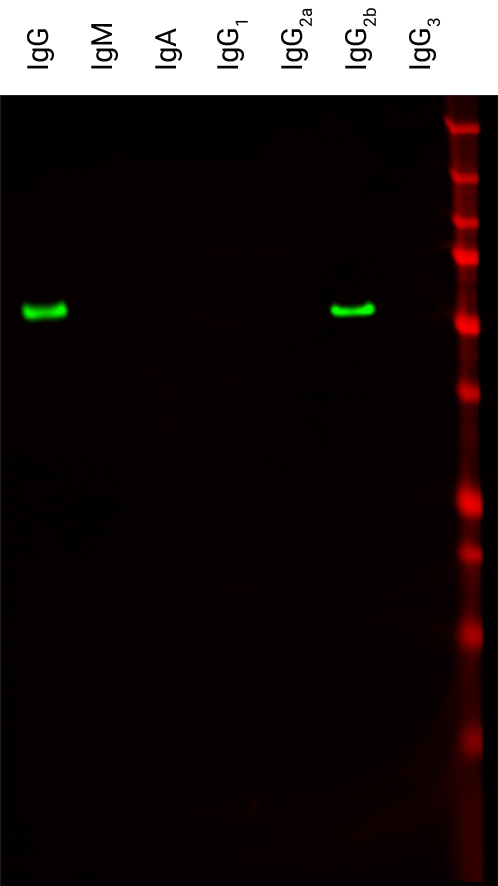
Purified mouse subclasses were detected by Western blot with IRDye 800CW Goat anti-Mouse IgG2b (P/N 926-32352) to demonstrate that it detects the heavy chains of mouse IgG and mouse IgG2b.

LI-COR IRDye® 800CW 山羊抗小鼠 IgM(μ 链特异性)二抗,LI-COR 二抗

Reagents > IRDye® 800CW Goat anti-Mouse IgM (µ chain specific) Secondary Antibody,IRDye® 800CW 山羊抗小鼠 IgM(μ 链特异性)二抗

IRDye® 800CW Goat anti-Mouse IgM (µ chain specific) Secondary Antibody

Immunogen

Mouse IgM paraproteins

Purity and Specificity

Isolation of specific antibodies was accomplished by affinity chromatography using mouse IgM covalently linked to agarose. Based on ELISA and/or flow cytometry, this antibody reacts with the heavy chain of mouse IgM. This antibody has been tested by dot blot and/or solid-phase adsorbed to ensure minimal cross-reactivity with mouse IgG1, IgG2a, IgG3, and IgA, pooled human sera and purified human paraproteins. The conjugate has been specifically tested and qualified for Western blot applications.

使用与琼脂糖共价连接的小鼠 IgM 通过亲和层析实现特异性抗体的分离。 基于 ELISA 和/或流式细胞术,该抗体与小鼠 IgM 的重链发生反应。 该抗体已通过斑点印迹和/或固相吸附测试,以确保与小鼠 IgG1、IgG2a、IgG3 和 IgA、合并的人血清和纯化的人副蛋白的交叉反应最小。 该偶联物经过专门测试,可用于蛋白质印迹应用。

Applications

Highly recommended for:

  • Western Blot
  • In-Cell Western™ Assay
  • On-Cell Western Assay
  • Protein Array
  • Immunohistochemistry
  • Small Animal Imaging
  • Microscopy
  • 2D Gel Detection
  • Tissue Section Imaging
  • Virus Titration Assay
  • 蛋白质印迹
    In-Cell Western™ 检测
    On-Cell Western 检测
    蛋白质阵列
    免疫组织化学
    小动物成像
    显微镜
    2D 凝胶检测
    组织切片成像
    病毒滴定法

Formulation

IRDye 800CW secondary antibodies are supplied as purified immunoglobulin conjugates, lyophilized in phosphate-buffered saline, pH 7.4. Protect from light. Store at 4 °C prior to reconstitution.

Each vial contains 10 mg/mL BSA (free of IgG and protease) as a stabilizer and 0.01% sodium azide as a preservative, after reconstitution. Concentration is 1.0 mg/mL when reconstituted as directed. Refer to the pack insert for details on reconstitution.

IRDye 800CW 二抗以纯化的免疫球蛋白偶联物形式提供,在 pH 7.4 的磷酸盐缓冲盐水中冻干。 避光。 在重组前储存在 4°C。

复溶后,每瓶含有 10 mg/mL BSA(不含 IgG 和蛋白酶)作为稳定剂和 0.01% 叠氮化钠作为防腐剂。 按照指示重新配制时,浓度为 1.0 mg/mL。 有关重组的详细信息,请参阅包装插页。

Recommended Dilutions

Application Recommended Suggested Range
Odyssey® Western blot detection 1:15,000 1:5,000 – 1:25,000
Other User optimized

Optimum dilutions will vary and should be determined empirically.

RRID

  • P/N 925-32280: RRID AB_2814916
  • P/N 926-32280: RRID AB_2814919

LI-COR IRDye® 800CW 山羊抗兔 IgG 二抗,LI-COR IRDye 800CW 二抗

Reagents > IRDye® 800CW Goat anti-Rabbit IgG Secondary Antibody,IRDye® 800CW 山羊抗兔 IgG 二抗

IRDye® 800CW Goat anti-Rabbit IgG Secondary Antibody

Immunogen

Rabbit IgG

Purity and Specificity

Isolation of specific antibodies was accomplished by affinity chromatography using pooled rabbit IgG covalently linked to agarose. Based on ELISA and flow cytometry, this antibody reacts with the heavy and light chains of rabbit IgG, and with the light chains of rabbit IgM and IgA. This antibody was tested by dot blot and and/or solid-phase adsorbed for minimal cross-reactivity with human, mouse, rat, sheep, and chicken serum proteins, but may cross-react with immunoglobulins from other species. The conjugate has been specifically tested and qualified for Western blot and In-Cell Western™ assay applications.

使用与琼脂糖共价连接的混合兔 IgG,通过亲和层析实现特异性抗体的分离。 基于 ELISA 和流式细胞术,该抗体与兔 IgG 的重链和轻链以及兔 IgM 和 IgA 的轻链反应。 通过斑点印迹和/或固相吸附测试该抗体与人、小鼠、大鼠、绵羊和鸡血清蛋白的交叉反应最小,但可能与来自其他物种的免疫球蛋白交叉反应。 该偶联物经过专门测试,可用于蛋白质印迹和 In-Cell Western™ 检测应用。

Applications

Highly recommended for:

  • Western Blot
  • In-Cell Western Assay
  • On-Cell Western Assay
  • Protein Array
  • Immunohistochemistry
  • Small Animal Imaging
  • Microscopy
  • 2D Gel Detection
  • Tissue Section Imaging
  • Virus Titration Assay
  • 蛋白质印迹
    细胞内西方检测
    On-Cell Western 检测
    蛋白质阵列
    免疫组织化学
    小动物成像
    显微镜
    2D 凝胶检测
    组织切片成像
    病毒滴定法

Formulation

IRDye 800CW secondary antibodies are supplied as purified immunoglobulin conjugates, lyophilized in phosphate-buffered saline, pH 7.4. Protect from light. Store at 4 °C prior to reconstitution.

Each vial contains 10 mg/mL BSA (free of IgG and protease) as a stabilizer and 0.01% sodium azide as a preservative, after reconstitution. Concentration is 1.0 mg/mL when reconstituted as directed. Refer to the pack insert for details on reconstitution.

IRDye 800CW 二抗以纯化的免疫球蛋白偶联物形式提供,在 pH 7.4 的磷酸盐缓冲盐水中冻干。 避光。 在重组前储存在 4°C。

复溶后,每瓶含有 10 mg/mL BSA(不含 IgG 和蛋白酶)作为稳定剂和 0.01% 叠氮化钠作为防腐剂。 按照指示重新配制时,浓度为 1.0 mg/mL。 有关重组的详细信息,请参阅包装插页。

Recommended Dilutions

Application Recommended Suggested Range
Odyssey® Western blot detection 1:15,000 1:5,000 – 1:25,000
In-Cell Western assay detection 1:200 1:200 – 1:1,200
Other User optimized

Optimum dilutions will vary and should be determined empirically.

RRID

  • P/N 925-32211: RRID AB_2651127
  • P/N 926-32211: RRID AB_621843

LI-COR IRDye® 800CW 山羊抗大鼠 IgG 二抗, LI-COR IRDye® 800CW 二抗

Reagents > IRDye® 800CW Goat anti-Rat IgG Secondary Antibody,IRDye® 800CW 山羊抗大鼠 IgG 二抗

IRDye® 800CW Goat anti-Rat IgG Secondary Antibody

Immunogen

Rat IgG, whole molecule

Purity and Specificity

The antibody was purified from antisera by immunoaffinity chromatography using antigens coupled to agarose beads. Based on immunoelectrophoresis and/or ELISA, this antibody reacts with whole molecule rat IgG, and with the light chains of other rat immunoglobulins. No antibody was detected against non-immunoglobulin serum proteins. This antibody has been tested by ELISA and and/or solid-phase adsorbed to ensure minimal cross-reactivity with mouse, bovine, horse, human, and rabbit serum proteins, but may cross-react with immunoglobulins from other species. The conjugate has been specifically tested and qualified for Western blot applications.

使用与琼脂糖珠偶联的抗原,通过免疫亲和层析从抗血清中纯化抗体。 基于免疫电泳和/或 ELISA,该抗体与全分子大鼠 IgG 以及其他大鼠免疫球蛋白的轻链发生反应。 未检测到针对非免疫球蛋白血清蛋白的抗体。 该抗体已通过 ELISA 和/或固相吸附测试,以确保与小鼠、牛、马、人类和兔血清蛋白的交叉反应最小,但可能与其他物种的免疫球蛋白交叉反应。 该偶联物经过专门测试,可用于蛋白质印迹应用。

Applications

Highly recommended for:

  • Western Blot
  • In-Cell Western™ Assay
  • On-Cell Western Assay
  • Protein Array
  • Immunohistochemistry
  • Small Animal Imaging
  • Microscopy
  • 2D Gel Detection
  • Tissue Section Imaging
  • Virus Titration Assay

Formulation

IRDye 800CW secondary antibodies are supplied as purified immunoglobulin conjugates, lyophilized in phosphate-buffered saline, pH 7.4. Protect from light. Store at 4 °C prior to reconstitution.

Each vial contains 10 mg/mL BSA (free of IgG and protease) as a stabilizer and 0.01% sodium azide as a preservative, after reconstitution. Concentration is 1.0 mg/mL when reconstituted as directed. Refer to the pack insert for details on reconstitution.

IRDye 800CW 二抗以纯化的免疫球蛋白偶联物形式提供,在 pH 7.4 的磷酸盐缓冲盐水中冻干。 避光。 在重组前储存在 4°C。

复溶后,每瓶含有 10 mg/mL BSA(不含 IgG 和蛋白酶)作为稳定剂和 0.01% 叠氮化钠作为防腐剂。 按照指示重新配制时,浓度为 1.0 mg/mL。 有关重组的详细信息,请参阅包装插页。

Recommended Dilutions

Application Recommended Suggested Range
Odyssey Western blot detection 1:15,000 1:5,000 – 1:25,000
Other User optimized

Optimum dilutions will vary and should be determined empirically.

RRID

  • P/N 925-32219: RRID AB_2721932
  • P/N 926-32219: RRID AB_1850025

Example Data

IRDye® 800CW Goat anti-Rat IgG Secondary Antibody
Western blot detection of tubulin in C32 lysate. C32 lysate (Santa Cruz P/N sc- 2205) was resolved on a 10% Tris-Glycine gel and transferred to Odyssey® nitrocellulose (P/N 926-31090). The membrane was probed with rat anti-tubulin (Novus Biologicals P/N NB600-506) followed by detection with IRDye 800CW Goat anti-Rat IgG (P/N 926-32219).
蛋白质印迹检测 C32 裂解物中的微管蛋白。 C32 裂解物 (Santa Cruz P/N sc-2205) 在 10% Tris-Glycine 凝胶上分离并转移到 Odyssey® 硝酸纤维素 (P/N 926-31090)。 用大鼠抗微管蛋白(Novus Biologicals P/N NB600-506)探测膜,然后用 IRDye 800CW 山羊抗大鼠 IgG(P/N 926-32219)检测。

LI-COR IRDye® 800CW 马来酰亚胺,LI-COR IRDye® 800CW

Reagents > IRDye® 800CW Maleimide,IRDye® 800CW 马来酰亚胺

IRDye® 800CW Maleimide

IRDye 800CW Maleimide provides the functionality for labeling molecules that contain free sulfhydryl (–SH) groups, such as cysteine residues in proteins, with IRDye 800CW. The reactive group of the IRDye 800CW Maleimide allows conjugation reactions to be performed very efficiently at physiological pH.

IRDye 800CW 马来酰亚胺提供了用 IRDye 800CW 标记含有游离巯基 (-SH) 基团的分子的功能,例如蛋白质中的半胱氨酸残基。 IRDye 800CW 马来酰亚胺的反应性基团允许在生理 pH 值下非常有效地进行偶联反应。

IRDye® 800CW Maleimide
Maleimide reacting with a thiol group.

Dye Structure

IRDye® 800CW Maleimide

Properties

  • Chemical Formula: C52H57N4Na3O16S4
  • Molecular Weight: 1191.25 g/mol
  • Exact Mass: 1190.23

Absorption and Emission Spectra

Solvent Ext. Coeff. (M-1cm-1) Absorption Maxima Emission Maxima
Methanol 300,000 778 nm 794 nm
Water 240,000 774 nm 789 nm
1X PBS 240,000 774 nm 789 nm
PBS: Methanol 270,000 777 nm 791 nm

Absorption and Emission Spectra in 1X PBS

IRDye® 800CW Maleimide

LI-COR IRDye® 800CW NHS 酯,LI-COR Biosciences IRDye 红外染料

Reagents > IRDye® 800CW NHS Ester,IRDye® 800CW NHS 酯

IRDye® 800CW NHS Ester

IRDye infrared dyes from LI-COR Biosciences are available for researchers developing applications using near-infrared (NIR) fluorescence. These unique dyes are used in a variety of NIR imaging applications including Western blotting, plate-based assays, protein arrays, tissue section imaging, and molecular activity measurements.

Standard NHS ester chemistry is used to produce custom probes labeled with LI-COR IRDye infrared dyes. The NHS ester reactive group provides the functionality for labeling primary and secondary amines, such as lysine residues in proteins.

LI-COR Biosciences 的 IRDye 红外染料可供研究人员使用近红外 (NIR) 荧光开发应用。 这些独特的染料用于各种 NIR 成像应用,包括蛋白质印迹、基于板的测定、蛋白质阵列、组织切片成像和分子活性测量。

标准 NHS 酯化学用于生产用 LI-COR IRDye 红外染料标记的定制探针。 NHS 酯反应基团提供了标记伯胺和仲胺的功能,例如蛋白质中的赖氨酸残基。

IRDye® 800CW NHS Ester
NHS Ester reacts with a primary aliphatic amine, such as lysine.
IRDye® 800CW NHS Ester
Dye label is attached through amide bond.

Dye Structure

IRDye® 800CW NHS Ester

Properties

  • Chemical Formula: C50H54N3Na3O17S4
  • Molecular Weight: 1166.20 g/mol
  • Exact Mass: 1165.20

Absorption and Emission Spectra

Solvent Ext. Coeff. (M-1cm-1) Absorption Maxima Emission Maxima
Methanol 300,000 778 nm 794 nm
Water 240,000 774 nm 789 nm
1X PBS 240,000 774 nm 789 nm
PBS: Methanol 270,000 777 nm 791 nm

Absorption and Emission Spectra in 1X PBS

IRDye® 800CW NHS Ester

LI-COR IRDye® 800CW 蛋白质标记试剂盒,LI-COR IRDye 800CW

Reagents > IRDye® 800CW Protein Labeling Kits,IRDye® 800CW 蛋白质标记试剂盒

IRDye® 800CW Protein Labeling Kits

IRDye 800CW Protein Labeling Kits can be used to label antibodies and other proteins for applications such as Western blots, In-Cell Western™ assays, in vivo imaging, and whole organ or tissue section assays.

IRDye 800CW 蛋白质标记试剂盒可用于标记抗体和其他蛋白质,用于蛋白质印迹、In-Cell Western™ 测定、体内成像以及整个器官或组织切片测定等应用。

High Molecular Weight

Optimized for labeling 1.0 mg amounts of protein with molecular weight 45 – 200 kDa.

针对标记 1.0 mg 分子量 45 – 200 kDa 的蛋白质进行了优化。

This kit contains:

    • 3 x 0.1 mg IRDye 800CW Reactive Dye vials
    • 0.5 mL 1 M Potassium Phosphate (K2HPO4), pH 9
    • 25 mL 1X PBS
    • 0.5 mL ultra-pure water
    • 3 x Pierce® Zeba™ Desalting Spin Columns (P/N 89891)

Note: The minimum recommended protein molecular weight for these columns is 7 kDa. See IRDye Peptide Labeling Guide for options.

  • Pierce Zeba Desalting Spin column instructions
  • Protocol for IRDye 800CW Protein Labeling Kit – High MW

Low Molecular Weight

Optimized for labeling 1.0 mg of protein with molecular weight 14 – 45 kDa.

This kit contains:

    • 3 x 0.5 mg IRDye 800CW Reactive Dye vials
    • 0.5 mL 1 M Potassium Phosphate (K2HPO4), pH 9
    • 25 mL 1X PBS
    • 0.5 mL ultra-pure water
    • 3 x Pierce Zeba Desalting Spin Columns (P/N 89891)

Note: The minimum recommended protein molecular weight for these columns is 7 kDa. See IRDye Peptide Labeling Guide for options.

  • Pierce Zeba Desalting Spin column instructions
  • Protocol for IRDye 800CW Protein Labeling Kit – Low MW

Microscale

Optimized for labeling 100 μg of protein with molecular weight 14 – 200 kDa.

This kit contains:

    • 3 x 0.1 mg IRDye 800CW Reactive Dye vials
    • 0.5 mL 1 M Potassium Phosphate (K2HPO4), pH 9
    • 25 mL 1X PBS
    • 0.5 mL ultra-pure water
    • 3 x Pierce Zeba Desalting Spin Columns (P/N 89892)

Note: The minimum recommended protein molecular weight for these columns is 7 kDa. See IRDye Peptide Labeling Guide for options.

  • Pierce Zeba Desalting Spin Column instructions
  • Protocol for IRDye 800CW Protein Labeling Kit – Microscale

LI-COR IRDye® 800CW RGD 光学探头,LI-COR IRDye® 800CW RGD

Reagents > IRDye® 800CW RGD Optical Probe,IRDye® 800CW RGD 光学探头

IRDye® 800CW RGD Optical Probe

IRDye 800CW RGD Optical Probe is a BrightSite™ near-infrared (NIR) fluorescently-labeled RGD imaging agent specifically designed to target the overexpression of integrins on tumors.

Integrins are cell surface heterodimeric glycoproteins involved in cell-to-cell and cell-to-matrix interactions. IRDye 800CW RGD Optical Probe is composed of the recognition motif, RGD (Arg-Gly-Asp), a tripeptide sequence used to bind integrin receptors including αvβ3. This receptor class is involved in tumor growth, tumor invasiveness, metastasis, tumor induced angiogenesis, inflammation, osteoporosis, and rheumatoid arthritis.

Interest in using a labeled RGD peptide ligand for the study and/or monitoring of diseases related to the αvβ3 receptor over-expression is increasing. Several groups have labeled RGD successfully with fluorescent dyes for in vitro and in vivo imaging.

IRDye 800CW RGD 光学探头是一种 BrightSite™ 近红外 (NIR) 荧光标记的 RGD 显像剂,专门设计用于靶向肿瘤上整合素的过度表达。

整联蛋白是细胞表面异二聚体糖蛋白,参与细胞与细胞和细胞与基质的相互作用。 IRDye 800CW RGD 光学探针由识别基序 RGD (Arg-Gly-Asp) 组成,这是一个三肽序列,用于结合包括 αvβ3 在内的整联蛋白受体。 该受体类别涉及肿瘤生长、肿瘤侵袭性、转移、肿瘤诱导的血管生成、炎症、骨质疏松症和类风湿性关节炎。

使用标记的 RGD 肽配体研究和/或监测与 αvβ3 受体过度表达相关的疾病的兴趣正在增加。 几个小组已经成功地用荧光染料标记了 RGD,用于体外和体内成像。

Learn about other LI-COR optical probes.

Applications

  • Microscopy
  • Optical Probe Development
  • Target Specificity

Absorbance and Emission Spectra

IRDye® 800CW RGD Optical Probe

Data Example

IRDye® 800CW RGD Optical Probe
A) The On-Cell Western cell-based assay was used to evaluate binding affinity of IRDye 800CW RGD to U87 (glioblastoma) and 22Rv1 (prostate carcinoma) tumor cell lines. B) Binding of the IRDye 800CW RGD peptide was blocked by unlabeled RGD but not by a non-specific RAD peptide in U87 tumor cells. Nude mice bearing subcutaneous tumors, U87 (left hip) and A431(right hip), were imaged 24 hours post intravenous injection of IRDye 800CW RGD (1 nmole; C) or IRDye 800CW RGD (1 nmole) following a pre-injection 1 hour prior to an injection of unlabeled RGD peptide (100 nmoles; D) Images were captured on the Pearl® Imager; 800 nm signal is presented in pseudo-color overlaid on a white light image of the mouse. The images are normalized to the same look-up-table.
A) 基于 On-Cell Western 细胞的测定用于评估 IRDye 800CW RGD 与 U87(胶质母细胞瘤)和 22Rv1(前列腺癌)肿瘤细胞系的结合亲和力。 B) IRDye 800CW RGD 肽的结合被未标记的 RGD 阻断,但未被 U87 肿瘤细胞中的非特异性 RAD 肽阻断。 在预注射 1 小时后静脉注射 IRDye 800CW RGD(1 nmole;C)或 IRDye 800CW RGD(1 nmole)后 24 小时对带有皮下肿瘤 U87(左髋)和 A431(右髋)的裸鼠进行成像 在注射未标记的 RGD 肽之前(100 nmoles;D)在 Pearl® Imager 上捕获图像; 800 nm 信号以覆盖在鼠标白光图像上的伪彩色呈现。 图像被标准化为相同的查找表。

Reference

  1. Huang, R., Vider, J., Kovar, J. L., Olive, D. M., Mellinghoff, I. K., Mayer-Kuckuk, P., … Blasberg, R. G. (2012). Integrin αvβ3-targeted IRDye 800CW near-infrared imaging of glioblastoma. Clinical Cancer Research, 18(20), 5731–5740. doi: 10.1158/1078-0432.CCR-12-0374.

LI-COR IRDye® 800CW 链霉亲和素,LI-COR IRDye® 800CW

Reagents > IRDye® 800CW Streptavidin,IRDye® 800CW 链霉亲和素

IRDye® 800CW Streptavidin

IRDye 800CW Streptavidin is supplied as a liquid in buffer containing 10 mM phosphate, 183 mM NaCl, 2.7 nM KCl, pH 7.4 with sodium azide 0.005% (w/v) as a preservative.

To use, centrifuge briefly before use to eliminate aggregates that may have formed in solution. This will reduce non-specific background staining. A final concentration of 0.2 to 1.0 µg/ml (1:1,000 to 1:5,000) is usually satisfactory for most applications; however, appropriate dilution may need to be determined empirically.

For membrane-based applications and In-Gel Westerns, it is recommended to add SDS (0.02% to 0.1% final concentration), in addition to Tween® 20 (0.1 to 0.2% final concentration) during the detection incubation step to reduce non-specific background staining.

IRDye 800CW 链霉亲和素以液体形式提供,缓冲液中含有 10 mM 磷酸盐、183 mM NaCl、2.7 nM KCl,pH 7.4,叠氮化钠 0.005% (w/v) 作为防腐剂。

要使用,请在使用前短暂离心以消除可能在溶液中形成的聚集体。 这将减少非特异性背景染色。 对于大多数应用,0.2 到 1.0 µg/ml(1:1,000 到 1:5,000)的最终浓度通常是令人满意的; 但是,可能需要根据经验确定适当的稀释度。

对于基于膜的应用和 In-Gel Westerns,建议在检测孵育步骤中添加 SDS(0.02% 至 0.1% 最终浓度)和 Tween® 20(0.1 至 0.2% 最终浓度)以减少非 特定背景染色。

Recommended Dilutions

Application Suggested Range Tween 20* SDS*
Odyssey® Western blot detection 1:1,000 – 1:5,000 0.1 – 0.2% (v/v) 0.02 – 0.1% (v/v)
Other User optimized User optimized User optimized

Optimum dilutions will vary and should be determined empirically.

* Added to reduce non-specific background staining.

See the complete line of IRDye Streptavidins.

LI-COR IRDye® 800RS NHS 酯,LI-COR IRDye 800RS 红外染料

Reagents > IRDye® 800RS NHS Ester,IRDye® 800RS NHS 酯

IRDye® 800RS NHS Ester

IRDye 800RS infrared dye is an excellent choice for nucleic acid applications. This near-infrared fluorescence dye has good water solubility, but low salt tolerance. It is more hydrophobic than IRDye 800CW.

IRDye 800RS NHS ester can be used to label the primary and secondary amino groups of DNA and RNA. Nucleic acids that have been labeled with IRDye 800RS can be easily purified by reverse-phase chromatography.

IRDye 800RS 红外染料是核酸应用的绝佳选择。 这种近红外荧光染料具有良好的水溶性,但耐盐性低。 它比 IRDye 800CW 更疏水。

IRDye 800RS NHS 酯可用于标记 DNA 和 RNA 的伯氨基和仲氨基。 用 IRDye 800RS 标记的核酸可以很容易地通过反相色谱法纯化。

IRDye Infrared Dyes Overview

Dye Structure

IRDye® 800RS NHS Ester

Properties

  • Chemical Formula: C50H56N3NaO11S2
  • Molecular Weight: 962.12 g/mol
  • Exact Mass: 961.33

Absorption and Emission Spectra

Solvent Ext. Coeff. (M-1cm-1) Absorption Maxima Emission Maxima Stokes Shift
Methanol 300,000 770 nm 786 nm 16 nm
Water 200,000 767 nm 786 nm 19 nm
1X PBS 200,000 767 nm 786 nm 19 nm
PBS: Methanol 270,000 770 nm 786 nm 16 nm

Absorption and Emission Spectra in 1X PBS

IRDye® 800RS NHS Ester

LI-COR IRDye® QC-1 NHS 酯,IRDye 800CW 配对用于光声

Reagents > IRDye® QC-1 NHS Ester,IRDye® QC-1 NHS 酯

IRDye® QC-1 NHS Ester

IRDye QC-1 is the first non-fluorescent (dark) quencher compatible with a wide range of visible and near-infrared fluorophores (~500-800 nm). It has the widest available quenching range, so you do not need to carefully match the donor’s fluorescence spectrum with the acceptor’s absorbance. IRDye QC-1 quenches common fluorophores with >97% efficiency1.

IRDye QC-1 是第一个与各种可见光和近红外荧光团(~500-800 nm)兼容的非荧光(暗)猝灭剂。 它具有最宽的可用猝灭范围,因此您无需仔细匹配供体的荧光光谱与受体的吸光度。 IRDye QC-1 以 >97% 的效率淬灭常见的荧光团1。

IRDye® QC-1 NHS Ester
Energy that is transferred from the donor (Dem) can be emitted at the acceptor’s wavelength (Aem), quenching fluorescence.

For more information, see “Spectral Overlap of IRDye QC-1 Absorption with Common Donor Fluorophores” below.

IRDye QC-1 NHS ester can be used for FRET (fluorescence resonance energy transfer) applications such as protease assays. Recently, it has been paired with IRDye 800CW in optoacoustic studies.

有关更多信息,请参阅下面的“IRDye QC-1 吸收与常见供体荧光团的光谱重叠”。

IRDye QC-1 NHS 酯可用于 FRET(荧光共振能量转移)应用,例如蛋白酶分析。 最近,它已与 IRDye 800CW 配对用于光声研究。

IRDye Infrared Dyes Overview

Dye Structure

IRDye® QC-1 NHS Ester

Properties

  • Chemical Formula: C53H62C|N4Na3O16S4
  • Molecular Weight: 1243.75 g/mol
  • Exact Mass: 1242.24

Absorption Spectrum

Solvent Ext. Coeff. (M-1cm-1) Absorption Maxima
1X PBS 96,000 737 nm
Water 98,000 788 nm

Absorption Spectrum in 1X PBS

IRDye® QC-1 NHS Ester

Absorption Spectrum in Methanol

IRDye® QC-1 NHS Ester

Spectral Overlap of IRDye QC-1 Absorption with Common Donor Fluorophores

IRDye® QC-1 NHS Ester
Normalized emission spectra for several common donor fluorophores, and the absorption spectrum of IRDye QC-1. All are quenched by IRDye QC-1 with >97% efficiency. Reprinted with permission from Analytical Biochemistry 388 (2009) 220-28.
IRDye® QC-1 NHS Ester
List of fluorophores and spectrally compatible quenchers (shaded box = compatible). Almost all of these common fluorophores are spectrally compatible with IRDye QC-1. No other quencher spans this wide range or is compatible with wavelengths near 800 nm. Reprinted with permission from Analytical Biochemistry 388 (2009) 220-28.

Reference

  1. Peng, X et al. A nonfluorescent, broad range quencher dye for Fõrster resonance energy transfer assays. Anal Biochem. 388(2):220-28 (2009).

LI-COR IRDye® 680LT 驴抗鸡二抗,LI-COR IRDye 680LT 二抗

Reagents > IRDye® 680LT Donkey anti-Chicken Secondary Antibody,IRDye® 680LT 驴抗鸡二抗

IRDye® 680LT Donkey anti-Chicken Secondary Antibody

Immunogen

Chicken IgY, whole molecule. IgY is the original designation for the IgG-like protein found in both serum and egg yolk.

Purity and Specificity

The antibody was isolated from antisera by immunoaffinity chromatography using antigens coupled to agarose beads. Based on immunoelectrophoresis and/or ELISA, the antibody reacts with whole molecule chicken IgY, and with the light chains of other chicken immunoglobulins. No reactivity was detected against non-immunoglobulin serum proteins. This antibody was tested by ELISA and/or solid-phase adsorbed to ensure minimal cross-reactivity with bovine, goat, guinea pig, Syrian hamster, horse, human, mouse, rabbit, rat, and sheep serum proteins, but may cross-react with immunoglobulins from other species. The conjugate has been specifically tested and qualified for Western blot applications.

使用与琼脂糖珠偶联的抗原,通过免疫亲和层析从抗血清中分离抗体。 基于免疫电泳和/或 ELISA,抗体与全分子鸡 IgY 以及其他鸡免疫球蛋白的轻链反应。 未检测到针对非免疫球蛋白血清蛋白的反应性。 该抗体通过 ELISA 和/或固相吸附测试,以确保与牛、山羊、豚鼠、叙利亚仓鼠、马、人、小鼠、兔、大鼠和绵羊血清蛋白的交叉反应最小,但可能发生交叉反应 与来自其他物种的免疫球蛋白。 该偶联物经过专门测试,可用于蛋白质印迹应用。

Applications

Recommended for:

  • Western Blot
  • Protein Array
  • Immunohistochemistry
  • Microscopy
  • 2D Gel Detection
  • Tissue Section Imaging
  • 蛋白质印迹
    蛋白质阵列
    免疫组织化学
    显微镜
    2D 凝胶检测
    组织切片成像

Not Recommended for:

  • Small Animal Imaging
  • In-Cell Western Assay
  • On-Cell Western Assay

Formulation

IRDye 680LT secondary antibodies are supplied as purified immunoglobulin conjugates, lyophilized in phosphate-buffered saline, pH 7.4. Protect from light. Store at 4 °C prior to reconstitution.

Each vial contains 10 mg/mL BSA (free of IgG and protease) as a stabilizer and 0.01% sodium azide as a preservative, after reconstitution. Concentration is 1.0 mg/mL when reconstituted as directed. Refer to the pack insert for details on reconstitution.

IRDye 680LT 二抗以纯化的免疫球蛋白偶联物形式提供,在磷酸盐缓冲盐水中冻干,pH 7.4。 避光。 在重组前储存在 4°C。

复溶后,每瓶含有 10 mg/mL BSA(不含 IgG 和蛋白酶)作为稳定剂和 0.01% 叠氮化钠作为防腐剂。 按照指示重新配制时,浓度为 1.0 mg/mL。 有关重组的详细信息,请参阅包装插页。

Recommended Dilutions

Application Recommended Suggested Range
Odyssey Western blot detection 1:20,000 1:20,000 – 1:50,000
Other User optimized

Optimum dilutions will vary and should be determined empirically.

Note: When using PVDF membranes for Western blotting applications, SDS (final concentration of 0.01 – 0.02%) and Tween® 20 (final concentration of 0.1 – 0.2%) must be added during the detection incubation step to avoid non-specific background staining.

RRID

  • P/N 925-68028: RRID AB_2814923
  • P/N 926-68028: RRID AB_10707008

Example Data

IRDye® 680LT Donkey anti-Chicken Secondary Antibody
Western blot analysis of PTEN expression in mouse PTEN transfected 293T whole cell lysate (lane 2) and non-transfected 293T lysate (lane 3). Both lysates were loaded with 2 µg total protein per lane. Odyssey® One-Color Molecular Weight Marker (P/N 928-40000) was loaded in lane 1. The blot was probed with mouse ant-PTEN (IgG2b) (Santa Cruz P/N sc-133197) and chicken anti-GAPDH (ProSci P/N XW-7214) followed by detection with IRDye 800CW Donkey anti-Mouse (P/N 926-32212) and IRDye 680LT Donkey anti-Chicken (P/N 926-68028).
小鼠 PTEN 转染的 293T 全细胞裂解物(泳道 2)和未转染的 293T 裂解物(泳道 3)中 PTEN 表达的蛋白质印迹分析。 两种裂解物每条泳道均加载 2 µg 总蛋白。 将 Odyssey® One-Color Molecular Weight Marker (P/N 928-40000) 加载到泳道 1。用小鼠 ant-PTEN (IgG2b) (Santa Cruz P/N sc-133197) 和鸡抗 GAPDH ( ProSci P/N XW-7214),然后使用 IRDye 800CW 驴防鼠 (P/N 926-32212) 和 IRDye 680LT 驴防鸡 (P/N 926-68028) 进行检测。

LI-COR IRDye® 680LT 驴抗山羊 IgG 二抗,LI-COR IRDye 680LT 二抗

Reagents > IRDye® 680LT Donkey anti-Goat IgG Secondary Antibody,IRDye® 680LT 驴抗山羊 IgG 二抗

IRDye® 680LT Donkey anti-Goat IgG Secondary Antibody

Immunogen

Goat IgG

Purity and Specificity

Isolation of specific antibodies was accomplished by affinity chromatography using goat IgG covalently linked to agarose. Based on ELISA, this antibody reacts with the heavy and light chains of goat IgG and sheep IgG. This antibody has been tested by dot blot and/or solid-phase adsorbed to ensure minimal cross-reactivity with human, mouse, rabbit, rat, chicken. guinea pig, hamster, swine, and horse serum proteins, but may cross-react with immunoglobulins from other species. The conjugate has been specifically tested and qualified for Western blot applications.

使用与琼脂糖共价连接的山羊 IgG 通过亲和层析实现特异性抗体的分离。 基于 ELISA,该抗体与山羊 IgG 和绵羊 IgG 的重链和轻链反应。 该抗体已通过斑点印迹和/或固相吸附测试,以确保与人、小鼠、兔、大鼠、鸡的交叉反应最小。 豚鼠、仓鼠、猪和马血清蛋白,但可能与其他物种的免疫球蛋白发生交叉反应。 该偶联物经过专门测试,可用于蛋白质印迹应用。

Applications

Recommended for:

  • Western Blot
  • Protein Array
  • Immunohistochemistry
  • Microscopy
  • 2D Gel Detection
  • Tissue Section Imaging

Not Recommended for:

  • Small Animal Imaging
  • In-Cell Western Assay
  • On-Cell Western Assay

Note: Do not use this secondary antibody in combination with any secondary antibody whose host is goat (for example, IRDye 800CW Goat anti-Rabbit) for two-color Western blot detection.

Formulation

IRDye 680LT secondary antibodies are supplied as purified immunoglobulin conjugates, lyophilized in phosphate-buffered saline, pH 7.4. Protect from light. Store at 4 °C prior to reconstitution.

Each vial contains 10 mg/mL BSA (free of IgG and protease) as a stabilizer and 0.01% sodium azide as a preservative, after reconstitution. Concentration is 1.0 mg/mL when reconstituted as directed. Refer to the pack insert for details on reconstitution.

IRDye 680LT 二抗以纯化的免疫球蛋白偶联物形式提供,在磷酸盐缓冲盐水中冻干,pH 7.4。 避光。 在重组前储存在 4°C。

复溶后,每瓶含有 10 mg/mL BSA(不含 IgG 和蛋白酶)作为稳定剂和 0.01% 叠氮化钠作为防腐剂。 按照指示重新配制时,浓度为 1.0 mg/mL。 有关重组的详细信息,请参阅包装插页。

Recommended Dilutions

Application Recommended Suggested Range
Odyssey Western blot detection 1:20,000 1:20,000 – 1:50,000
Other User optimized

Optimum dilutions will vary and should be determined empirically.

Note: When using PVDF membranes for Western blotting applications, SDS (final concentration of 0.01 – 0.02%) and Tween® 20 (final concentration of 0.1 – 0.2%) must be added during the detection incubation step to avoid non-specific background staining.

RRID

  • P/N 925-68024: RRID AB_2814908
  • P/N 926-68024: RRID AB_10706168

Example Data

IRDye® 680LT Donkey anti-Goat IgG Secondary Antibody
Western blot detection of actin in C32 lysate. C32 lysate (Santa Cruz P/N sc- 2205) was resolved on a 10% Bis-Tris gel and transferred to Odyssey® nitrocellulose (P/N 926-31090). The membrane was probed with goat anti-actin (Santa Cruz P/N sc-1616) followed by detection with IRDye 680LT Donkey anti-Goat IgG (P/N 926-68024).
蛋白质印迹检测 C32 裂解物中的肌动蛋白。 C32 裂解物 (Santa Cruz P/N sc-2205) 在 10% Bis-Tris 凝胶上分离并转移到 Odyssey® 硝酸纤维素 (P/N 926-31090)。 用山羊抗肌动蛋白 (Santa Cruz P/N sc-1616) 探测膜,然后用 IRDye 680LT 驴抗山羊 IgG (P/N 926-68024) 检测。

LI-COR IRDye® 680LT 驴抗小鼠 IgG 二抗,LI-COR IRDye 680LT 二抗

Reagents > IRDye® 680LT Donkey anti-Mouse IgG Secondary Antibody,IRDye® 680LT 驴抗小鼠 IgG 二抗

IRDye® 680LT Donkey anti-Mouse IgG Secondary Antibody

Immunogen

Mouse IgG

Purity and Specificity

The antibody was isolated by affinity chromatography using antigens coupled to agarose beads. Based on ELISA, this antibody reacts with the heavy and light chains of mouse IgG, and with the light chains of mouse IgM and IgA. This antibody was tested by ELISA and/or solid-phase adsorbed to ensure minimal cross-reactivity with bovine, chicken, goat, guinea pig, horse, human, rabbit, and sheep serum proteins, but may cross-react with immunoglobulins from other species. The conjugate has been specifically tested and qualified for Western blot applications.

使用与琼脂糖珠偶联的抗原通过亲和层析分离抗体。 基于 ELISA,该抗体与小鼠 IgG 的重链和轻链以及小鼠 IgM 和 IgA 的轻链反应。 该抗体通过 ELISA 和/或固相吸附测试,以确保与牛、鸡、山羊、豚鼠、马、人、兔和绵羊血清蛋白的交叉反应最小,但可能与来自其他物种的免疫球蛋白交叉反应 . 该偶联物经过专门测试,可用于蛋白质印迹应用。

Applications

Recommended for:

  • Western Blot
  • Protein Array
  • Immunohistochemistry
  • Microscopy
  • 2D Gel Detection
  • Tissue Section Imaging
  • 蛋白质印迹
    蛋白质阵列
    免疫组织化学
    显微镜
    2D 凝胶检测
    组织切片成像

Not Recommended for:

  • Small Animal Imaging
  • In-Cell Western Assay
  • On-Cell Western Assay
Note: IRDye 680LT secondary antibodies are not intended for In-Cell Western™ assays or in vivo imaging applications.

Formulation

IRDye 680LT secondary antibodies are supplied as purified immunoglobulin conjugates, lyophilized in phosphate-buffered saline, pH 7.4. Protect from light. Store at 4 °C prior to reconstitution.

Each vial contains 10 mg/mL BSA (free of IgG and protease) as a stabilizer and 0.01% sodium azide as a preservative, after reconstitution. Concentration is 1.0 mg/mL when reconstituted as directed. Refer to the pack insert for details on reconstitution.

IRDye 680LT 二抗以纯化的免疫球蛋白偶联物形式提供,在磷酸盐缓冲盐水中冻干,pH 7.4。 避光。 在重组前储存在 4°C。

复溶后,每瓶含有 10 mg/mL BSA(不含 IgG 和蛋白酶)作为稳定剂和 0.01% 叠氮化钠作为防腐剂。 按照指示重新配制时,浓度为 1.0 mg/mL。 有关重组的详细信息,请参阅包装插页。

Recommended Dilutions

Application Recommended Suggested Range
Odyssey Western blot detection 1:20,000 1:20,000 – 1:50,000
Other User optimized

Optimum dilutions will vary and should be determined empirically.

Note: When using PVDF membranes for Western blotting applications, SDS (final concentration of 0.01 – 0.02%) and Tween® 20 (final concentration of 0.1 – 0.2%) must be added during the detection incubation step to avoid non-specific background staining.

RRID

  • P/N 925-68022: RRID AB_2814906
  • P/N 926-68022: RRID AB_10715072

Example Data

IRDye® 680LT Donkey anti-Mouse IgG Secondary Antibody
Western blot detection of actin in C32 lysate. Samples loaded in 4X Protein Sample Loading Buffer (P/N 928-40004), resolved on a 10% Bis-Tris gel, and transferred to Odyssey® nitrocellulose (P/N 926-31090). The membrane was probed with mouse anti-actin (Neomarkers P/N MS-1295) followed by detection with IRDye 680LT Donkey anti-Mouse IgG (P/N 926-68022).

LI-COR IRDye® 680LT 驴抗兔 IgG 二抗,LI-COR IRDye 680LT 二抗

Reagents > IRDye® 680LT Donkey anti-Rabbit IgG Secondary Antibody,IRDye® 680LT 驴抗兔 IgG 二抗

IRDye® 680LT Donkey anti-Rabbit IgG Secondary Antibody

Immunogen

Rabbit IgG, whole molecule

Purity and Specificity

The antibody was isolated from antisera by immunoaffinity chromatography using antigens coupled to agarose beads. Based on immunoelectrophoresis, this antibody reacts with the heavy chains of rabbit IgG, and with the light chains common to most rabbit immunoglobulins. No reactivity was detected against non-immunoglobulin serum proteins. This antibody was tested by ELISA and/or solid-phase adsorbed to ensure minimal cross-reactivity with bovine, chicken, goat, guinea pig, Syrian hamster, horse, human, mouse, rat, and sheep serum proteins, but may cross-react with immunoglobulins from other species. The conjugate has been specifically tested and qualified for Western blot applications.

使用与琼脂糖珠偶联的抗原,通过免疫亲和层析从抗血清中分离抗体。 基于免疫电泳,该抗体与兔 IgG 的重链以及大多数兔免疫球蛋白常见的轻链反应。 未检测到针对非免疫球蛋白血清蛋白的反应性。 该抗体经过 ELISA 和/或固相吸附测试,以确保与牛、鸡、山羊、豚鼠、叙利亚仓鼠、马、人、小鼠、大鼠和绵羊血清蛋白的交叉反应最小,但可能发生交叉反应 与来自其他物种的免疫球蛋白。 该偶联物经过专门测试,可用于蛋白质印迹应用。

Applications

Recommended for:

  • Western Blot
  • Protein Array
  • Immunohistochemistry
  • Microscopy
  • 2D Gel Detection
  • Tissue Section Imaging

Not Recommended for:

  • Small Animal Imaging
  • In-Cell Western Assay
  • On-Cell Western Assay

Formulation

IRDye 680LT secondary antibodies are supplied as purified immunoglobulin conjugates, lyophilized in phosphate-buffered saline, pH 7.4. Protect from light. Store at 4 °C prior to reconstitution.

Each vial contains 10 mg/mL BSA (free of IgG and protease) as a stabilizer and 0.01% sodium azide as a preservative, after reconstitution. Concentration is 1.0 mg/mL when reconstituted as directed. Refer to the pack insert for details on reconstitution.

IRDye 680LT 二抗以纯化的免疫球蛋白偶联物形式提供,在磷酸盐缓冲盐水中冻干,pH 7.4。 避光。 在重组前储存在 4°C。

复溶后,每瓶含有 10 mg/mL BSA(不含 IgG 和蛋白酶)作为稳定剂和 0.01% 叠氮化钠作为防腐剂。 按照指示重新配制时,浓度为 1.0 mg/mL。 有关重组的详细信息,请参阅包装插页。

Recommended Dilutions

Application Recommended Suggested Range
Odyssey Western blot detection 1:20,000 1:20,000 – 1:50,000
Other User optimized

Optimum dilutions will vary and should be determined empirically.

Note: When using PVDF membranes for Western blotting applications, SDS (final concentration of 0.01 – 0.02%) and Tween® 20 (final concentration of 0.1 – 0.2%) must be added during the detection incubation step to avoid non-specific background staining.

RRID

  • P/N 925-68023: RRID AB_2814907
  • P/N 926-68023: RRID AB_10706167

LI-COR IRDye® 680LT 山羊抗人 IgG 二抗,LI-COR IRDye 680LT 二抗

Reagents > IRDye® 680LT Goat anti-Human IgG Secondary Antibody,IRDye® 680LT 山羊抗人 IgG 二抗

IRDye® 680LT Goat anti-Human IgG Secondary Antibody

Immunogen

Human IgG

Purity and Specificity

Isolation of specific antibodies was accomplished by affinity chromatography using human IgG covalently linked to agarose. Based on ELISA, this antibody reacts with the heavy and light chains of human IgG. This antibody was tested by dot blot and/or solid-phase adsorbed to ensure minimal cross-reactivity with bovine, horse, and mouse serum proteins, but may cross-react with immunoglobulins from other species. The conjugate has been specifically tested and qualified for Western blot applications.

使用与琼脂糖共价连接的人 IgG 通过亲和层析实现特异性抗体的分离。 基于 ELISA,该抗体与人 IgG 的重链和轻链反应。 该抗体通过斑点印迹和/或固相吸附测试,以确保与牛、马和小鼠血清蛋白的交叉反应最小,但可能与来自其他物种的免疫球蛋白交叉反应。 该偶联物经过专门测试,可用于蛋白质印迹应用。

Applications

Recommended for:

  • Western Blot
  • Protein Array
  • Immunohistochemistry
  • Microscopy
  • 2D Gel Detection
  • Tissue Section Imaging
  • 蛋白质印迹
    蛋白质阵列
    免疫组织化学
    显微镜
    2D 凝胶检测
    组织切片成像

Not Recommended for:

  • Small Animal Imaging
  • In-Cell Western Assay
  • On-Cell Western Assay

Formulation

IRDye 680LT secondary antibodies are supplied as purified immunoglobulin conjugates, lyophilized in phosphate-buffered saline, pH 7.4. Protect from light. Store at 4 °C prior to reconstitution.

Each vial contains 10 mg/mL BSA (free of IgG and protease) as a stabilizer and 0.01% sodium azide as a preservative, after reconstitution. Concentration is 1.0 mg/mL when reconstituted as directed. Refer to the pack insert for details on reconstitution.

IRDye 680LT 二抗以纯化的免疫球蛋白偶联物形式提供,在磷酸盐缓冲盐水中冻干,pH 7.4。 避光。 在重组前储存在 4°C。

复溶后,每瓶含有 10 mg/mL BSA(不含 IgG 和蛋白酶)作为稳定剂和 0.01% 叠氮化钠作为防腐剂。 按照指示重新配制时,浓度为 1.0 mg/mL。 有关重组的详细信息,请参阅包装插页。

Recommended Dilutions

Application Recommended Suggested Range
Odyssey Western blot detection 1:20,000 1:20,000 – 1:50,000
Other User optimized

Optimum dilutions will vary and should be determined empirically.

Note: When using PVDF membranes for Western blotting applications, SDS (final concentration of 0.01 – 0.02%) and Tween® 20 (final concentration of 0.1 – 0.2%) must be added during the detection incubation step to avoid non-specific background staining.

RRID

  • P/N 925-68032: RRID AB_2814911
  • P/N 926-68032: RRID AB_10795013

LI-COR IRDye® 680LT 山羊抗小鼠 IgG 二抗,LI-COR IRDye 680LT 二抗

Reagents > IRDye® 680LT Goat anti-Mouse IgG Secondary Antibody,IRDye® 680LT 山羊抗小鼠 IgG 二抗

IRDye® 680LT Goat anti-Mouse IgG Secondary Antibody

Immunogen

Mouse IgG paraproteins

Purity and Specificity

Isolation of specific antibodies was accomplished by affinity chromatography using pooled mouse IgG covalently linked to agarose. Based on ELISA and flow cytometry, this antibody reacts with the heavy and light chains of mouse IgG1, IgG2a, IgG2b, and IgG3, and with the light chains of mouse IgM and IgA. This antibody was tested by dot blot and and/or solid-phase adsorbed for minimal cross-reactivity with human, rabbit, goat, rat, and horse serum proteins, but may cross-react with immunoglobulins from other species. The conjugate has been specifically tested and qualified for Western blot applications.

使用与琼脂糖共价连接的混合小鼠 IgG 通过亲和层析实现特异性抗体的分离。 基于 ELISA 和流式细胞术,该抗体与小鼠 IgG1、IgG2a、IgG2b 和 IgG3 的重链和轻链以及小鼠 IgM 和 IgA 的轻链反应。 通过斑点印迹和/或固相吸附测试该抗体与人、兔、山羊、大鼠和马血清蛋白的交叉反应最小,但可能与来自其他物种的免疫球蛋白交叉反应。 该偶联物经过专门测试,可用于蛋白质印迹应用。

Applications

Recommended for:

  • Western Blot
  • Protein Array
  • Immunohistochemistry
  • Microscopy
  • 2D Gel Detection
  • Tissue Section Imaging
  • 蛋白质印迹
    蛋白质阵列
    免疫组织化学
    显微镜
    2D 凝胶检测
    组织切片成像

Not Recommended for:

  • Small Animal Imaging
  • In-Cell Western Assay
  • On-Cell Western Assay

Formulation

IRDye 680LT secondary antibodies are supplied as purified immunoglobulin conjugates, lyophilized in phosphate-buffered saline, pH 7.4. Protect from light. Store at 4 °C prior to reconstitution.

Each vial contains 10 mg/mL BSA (free of IgG and protease) as a stabilizer and 0.01% sodium azide as a preservative, after reconstitution. Concentration is 1.0 mg/mL when reconstituted as directed. Refer to the pack insert for details on reconstitution.

IRDye 680LT 二抗以纯化的免疫球蛋白偶联物形式提供,在磷酸盐缓冲盐水中冻干,pH 7.4。 避光。 在重组前储存在 4°C。

复溶后,每瓶含有 10 mg/mL BSA(不含 IgG 和蛋白酶)作为稳定剂和 0.01% 叠氮化钠作为防腐剂。 按照指示重新配制时,浓度为 1.0 mg/mL。 有关重组的详细信息,请参阅包装插页。

Recommended Dilutions

Application Recommended Suggested Range
Odyssey Western blot detection 1:20,000 1:20,000 – 1:50,000
Other User optimized

Optimum dilutions will vary and should be determined empirically.

Note: When using PVDF membranes for Western blotting applications, SDS (final concentration of 0.01 – 0.02%) and Tween® 20 (final concentration of 0.1 – 0.2%) must be added during the detection incubation step to avoid non-specific background staining.

RRID

  • P/N 925-68020: RRID AB_2687826
  • P/N 926-68020: RRID AB_10706161

Example Data

IRDye® 680LT Goat anti-Mouse IgG Secondary Antibody
Western blot detection of ERK2 in Hela lysate (5 µg – 78 ng). Samples loaded in 4X protein loading buffer (P/N 928-40004), resolved on a 10% Bis-Tris gel, and transferred to Odyssey® nitrocellulose (P/N 926-31090). The membrane was probed with mouse anti-ERK2 (Santa Cruz P/N sc-1647) followed by detection with IRDye 680LT Goat anti-Mouse IgG (P/N 926-68020).
对 Hela 裂解液 (5 µg – 78 ng) 中的 ERK2 进行蛋白质印迹检测。 样品上样于 4X 蛋白上样缓冲液 (P/N 928-40004),在 10% Bis-Tris 凝胶上分离,然后转移至 Odyssey® 硝酸纤维素 (P/N 926-31090)。 用小鼠抗 ERK2 (Santa Cruz P/N sc-1647) 探测膜,然后用 IRDye 680LT 山羊抗小鼠 IgG (P/N 926-68020) 检测。

LI-COR IRDye 680LT 二抗,IRDye® 680LT 山羊抗小鼠 IgM(μ 链特异性)二抗

Reagents > IRDye® 680LT Goat anti-Mouse IgM (µ chain specific) Secondary Antibody,IRDye® 680LT 山羊抗小鼠 IgM(μ 链特异性)二抗

IRDye® 680LT Goat anti-Mouse IgM (µ chain specific) Secondary Antibody

Immunogen

Mouse IgM paraproteins

Purity and Specificity

Isolation of specific antibodies was accomplished by affinity chromatography using mouse IgM covalently linked to agarose. Based on ELISA and/or flow cytometry, this antibody reacts with the heavy chain of mouse IgM. This antibody has been tested by dot blot and/or solid-phase adsorbed to ensure minimal cross-reactivity with mouse IgG1, IgG2a, IgG3, and IgA, pooled human sera and purified human paraproteins. The conjugate has been specifically tested and qualified for Western blot applications.

使用与琼脂糖共价连接的小鼠 IgM 通过亲和层析实现特异性抗体的分离。 基于 ELISA 和/或流式细胞术,该抗体与小鼠 IgM 的重链发生反应。 该抗体已通过斑点印迹和/或固相吸附测试,以确保与小鼠 IgG1、IgG2a、IgG3 和 IgA、合并的人血清和纯化的人副蛋白的交叉反应最小。 该偶联物经过专门测试,可用于蛋白质印迹应用。

Applications

Recommended for:

  • Western Blot
  • Protein Array
  • Immunohistochemistry
  • Microscopy
  • 2D Gel Detection
  • Tissue Section Imaging
  • 蛋白质印迹
    蛋白质阵列
    免疫组织化学
    显微镜
    2D 凝胶检测
    组织切片成像

Not Recommended for:

  • Small Animal Imaging
  • In-Cell Western Assay
  • On-Cell Western Assay

Formulation

IRDye 680LT secondary antibodies are supplied as purified immunoglobulin conjugates, lyophilized in phosphate-buffered saline, pH 7.4. Protect from light. Store at 4 °C prior to reconstitution.

Each vial contains 10 mg/mL BSA (free of IgG and protease) as a stabilizer and 0.01% sodium azide as a preservative, after reconstitution. Concentration is 1.0 mg/mL when reconstituted as directed. Refer to the pack insert for details on reconstitution.

IRDye 680LT 二抗以纯化的免疫球蛋白偶联物形式提供,在磷酸盐缓冲盐水中冻干,pH 7.4。 避光。 在重组前储存在 4°C。

复溶后,每瓶含有 10 mg/mL BSA(不含 IgG 和蛋白酶)作为稳定剂和 0.01% 叠氮化钠作为防腐剂。 按照指示重新配制时,浓度为 1.0 mg/mL。 有关重组的详细信息,请参阅包装插页。

Recommended Dilutions

Application Recommended Suggested Range
Odyssey Western blot detection 1:20,000 1:20,000 – 1:50,000
Other User optimized

Optimum dilutions will vary and should be determined empirically.

Note: When using PVDF membranes for Western blotting applications, SDS (final concentration of 0.01 – 0.02%) and Tween® 20 (final concentration of 0.1 – 0.2%) must be added during the detection incubation step to avoid non-specific background staining.
注意:当使用 PVDF 膜进行蛋白质印迹应用时,必须在检测孵育步骤中添加 SDS(终浓度为 0.01 – 0.02%)和 Tween® 20(终浓度为 0.1 – 0.2%)以避免非特异性背景染色。

RRID

  • P/N 925-68080: RRID AB_2814917
  • P/N 926-68080: RRID AB_2814920

Example Data

IRDye® 680LT Goat anti-Mouse IgM (µ chain specific) Secondary Antibody
Western blot detection of guinea pig IgG with IRDye 680LT Donkey anti-Guinea Pig. Serial dilutions of guinea pig IgG were spiked into C32 lysate (Santa Cruz P/N sc-2205). Samples were resolved on a 10% Bis-Tris gel and transferred to Odyssey® Nitrocellulose membrane (P/N 926-31092). The membrane was blocked with Odyssey Blocking Buffer (P/N 927-40000) followed by detection with IRDye 680LT Donkey anti-Guinea Pig (P/N 926-68030).
用 IRDye 680LT Donkey anti-Guinea Pig 对豚鼠 IgG 进行蛋白质印迹检测。 将连续稀释的豚鼠 IgG 加入 C32 裂解液 (Santa Cruz P/N sc-2205)。 样品在 10% Bis-Tris 凝胶上分离并转移到 Odyssey® 硝酸纤维素膜(P/N 926-31092)上。 用 Odyssey 封闭缓冲液 (P/N 927-40000) 封闭膜,然后用 IRDye 680LT 驴抗豚鼠 (P/N 926-68030) 检测。

LI-COR IRDye® 680LT 山羊抗兔 IgG 二抗,LI-COR IRDye 680LT 二抗

Reagents > IRDye® 680LT Goat anti-Rabbit IgG Secondary Antibody,IRDye® 680LT 山羊抗兔 IgG 二抗

IRDye® 680LT Goat anti-Rabbit IgG Secondary Antibody

Immunogen

Rabbit IgG

Purity and Specificity

Isolation of specific antibodies was accomplished by affinity chromatography using pooled rabbit IgG covalently linked to agarose. Based on ELISA and flow cytometry, this antibody reacts with the heavy and light chains of rabbit IgG, and with the light chains of rabbit IgM and IgA. This antibody was tested by dot blot and and/or solid-phase adsorbed for minimal cross-reactivity with human, mouse, rat, sheep, and chicken serum proteins, but may cross-react with immunoglobulins from other species. The conjugate has been specifically tested and qualified for Western blot applications.

使用与琼脂糖共价连接的混合兔 IgG,通过亲和层析实现特异性抗体的分离。 基于 ELISA 和流式细胞术,该抗体与兔 IgG 的重链和轻链以及兔 IgM 和 IgA 的轻链反应。 通过斑点印迹和/或固相吸附测试该抗体与人、小鼠、大鼠、绵羊和鸡血清蛋白的交叉反应最小,但可能与来自其他物种的免疫球蛋白交叉反应。 该偶联物经过专门测试,可用于蛋白质印迹应用。

Applications

Recommended for:

  • Western Blot
  • Protein Array
  • Immunohistochemistry
  • Microscopy
  • 2D Gel Detection
  • Tissue Section Imaging
  • 蛋白质印迹
    蛋白质阵列
    免疫组织化学
    显微镜
    2D 凝胶检测
    组织切片成像

Not Recommended for:

  • Small Animal Imaging
  • In-Cell Western Assay
  • On-Cell Western Assay

Formulation

IRDye 680LT secondary antibodies are supplied as purified immunoglobulin conjugates, lyophilized in phosphate-buffered saline, pH 7.4. Protect from light. Store at 4 °C prior to reconstitution.

Each vial contains 10 mg/mL BSA (free of IgG and protease) as a stabilizer and 0.01% sodium azide as a preservative, after reconstitution. Concentration is 1.0 mg/mL when reconstituted as directed. Refer to the pack insert for details on reconstitution.

IRDye 680LT 二抗以纯化的免疫球蛋白偶联物形式提供,在磷酸盐缓冲盐水中冻干,pH 7.4。 避光。 在重组前储存在 4°C。

复溶后,每瓶含有 10 mg/mL BSA(不含 IgG 和蛋白酶)作为稳定剂和 0.01% 叠氮化钠作为防腐剂。 按照指示重新配制时,浓度为 1.0 mg/mL。 有关重组的详细信息,请参阅包装插页。

Recommended Dilutions

Application Recommended Suggested Range
Odyssey Western blot detection 1:20,000 1:20,000 – 1:50,000
Other User optimized

Optimum dilutions will vary and should be determined empirically.

Note: When using PVDF membranes for Western blotting applications, SDS (final concentration of 0.01 – 0.02%) and Tween® 20 (final concentration of 0.1 – 0.2%) must be added during the detection incubation step to avoid non-specific background staining.
注意:当使用 PVDF 膜进行蛋白质印迹应用时,必须在检测孵育步骤中添加 SDS(终浓度为 0.01 – 0.02%)和 Tween® 20(终浓度为 0.1 – 0.2%)以避免非特异性背景染色。

RRID

  • P/N 925-68021: RRID AB_2713919
  • P/N 926-68021: RRID AB_10706309

Example Data

IRDye® 680LT Goat anti-Rabbit IgG Secondary Antibody
Western blot detection of p38 in Jurkat lysate. Samples loaded in 4X Protein Sample Loading Buffer (P/N 928-40004), resolved on a 10% Bis-Tris gel and transferred to Odyssey® nitrocellulose (P/N 926-31090). The membrane was blocked with Odyssey Blocking Buffer (P/N 927-40000) and probed with rabbit anti-p38 (Santa Cruz P/N sc-7149) followed by detection with IRDye 680LT Goat anti-Rabbit IgG (P/N 926-68021).
Jurkat 裂解物中 p38 的蛋白质印迹检测。 将样品装入 4X 蛋白质样品上样缓冲液 (P/N 928-40004),在 10% Bis-Tris 凝胶上分离,然后转移到 Odyssey® 硝酸纤维素 (P/N 926-31090)。 用 Odyssey 封闭缓冲液 (P/N 927-40000) 封闭膜,并用兔抗 p38 (Santa Cruz P/N sc-7149) 探测,然后用 IRDye 680LT 山羊抗兔 IgG (P/N 926- 68021)。

LI-COR IRDye® 680LT 山羊抗大鼠 IgG 二抗,LI-COR IRDye 680LT 二抗

Reagents > IRDye® 680LT Goat anti-Rat IgG Secondary Antibody,IRDye® 680LT 山羊抗大鼠 IgG 二抗

IRDye® 680LT Goat anti-Rat IgG Secondary Antibody

Immunogen

Rat IgG, whole molecule

Purity and Specificity

The antibody was purified from antisera by immunoaffinity chromatography using antigens coupled to agarose beads. Based on immunoelectrophoresis and/or ELISA, this antibody reacts with whole molecule rat IgG, and with the light chains of other rat immunoglobulins. No reactivity was detected against non-immunoglobulin serum proteins. This antibody has been tested by ELISA and and/or solid-phase adsorbed to ensure minimal cross-reactivity with mouse, bovine, horse, human, and rabbit serum proteins, but may cross-react with immunoglobulins from other species. The conjugate has been specifically tested and qualified for Western blot applications.

使用与琼脂糖珠偶联的抗原,通过免疫亲和层析从抗血清中纯化抗体。 基于免疫电泳和/或 ELISA,该抗体与全分子大鼠 IgG 以及其他大鼠免疫球蛋白的轻链发生反应。 未检测到针对非免疫球蛋白血清蛋白的反应性。 该抗体已通过 ELISA 和/或固相吸附测试,以确保与小鼠、牛、马、人类和兔血清蛋白的交叉反应最小,但可能与其他物种的免疫球蛋白交叉反应。 该偶联物经过专门测试,可用于蛋白质印迹应用。

Applications

Recommended for:

  • Western Blot
  • Protein Array
  • Immunohistochemistry
  • Microscopy
  • 2D Gel Detection
  • Tissue Section Imaging
  • 蛋白质印迹
    蛋白质阵列
    免疫组织化学
    显微镜
    2D 凝胶检测
    组织切片成像

Not Recommended for:

  • Small Animal Imaging
  • In-Cell Western Assay
  • On-Cell Western Assay

Formulation

IRDye 680LT secondary antibodies are supplied as purified immunoglobulin conjugates, lyophilized in phosphate-buffered saline, pH 7.4. Protect from light. Store at 4 °C prior to reconstitution.

Each vial contains 10 mg/mL BSA (free of IgG and protease) as a stabilizer and 0.01% sodium azide as a preservative, after reconstitution. Concentration is 1.0 mg/mL when reconstituted as directed. Refer to the pack insert for details on reconstitution.

IRDye 680LT 二抗以纯化的免疫球蛋白偶联物形式提供,在磷酸盐缓冲盐水中冻干,pH 7.4。 避光。 在重组前储存在 4°C。

复溶后,每瓶含有 10 mg/mL BSA(不含 IgG 和蛋白酶)作为稳定剂和 0.01% 叠氮化钠作为防腐剂。 按照指示重新配制时,浓度为 1.0 mg/mL。 有关重组的详细信息,请参阅包装插页。

Recommended Dilutions

Application Recommended Suggested Range
Odyssey Western blot detection 1:20,000 1:20,000 – 1:50,000
Other User optimized

Optimum dilutions will vary and should be determined empirically.

Note: When using PVDF membranes for Western blotting applications, SDS (final concentration of 0.01 – 0.02%) and Tween® 20 (final concentration of 0.1 – 0.2%) must be added during the detection incubation step to avoid non-specific background staining.

RRID

  • P/N 925-68029: RRID AB_2814909
  • P/N 926-68029: RRID AB_10715073

Example Data

IRDye® 680LT Goat anti-Rat IgG Secondary Antibody
Western blot detection of tubulin in C32 lysate. Samples loaded in 4X Protein Sample Loading Buffer (P/N 928-40004), resolved on a 10% Bis-Tris gel, and transferred to Odyssey® nitrocellulose (P/N 926-31090). The membrane was blocked with Odyssey Blocking Buffer (P/N 927-40000) and probed with rat anti-tubulin (Novus Biologicals P/N NB600-506) followed by detection with IRDye 680LT Goat anti-Rat IgG (P/N 926-68029).

LI-COR IRDye® 680RD BoneTag™ 光学探头,LI-COR IRDye 680RD 近红外染料

Reagents > IRDye® 680RD BoneTag™ Optical Probe

IRDye® 680RD BoneTag™ Optical Probe

IRDye 680RD BoneTag Optical Probe is a calcium-chelating compound conjugated to IRDye 680RD near-infrared dye. Using IRDye NIR dyes extends fluorescence signal detection to the near-infrared fluorescent region of the spectrum without affecting the compound’s ability to function as a marker of the mineralization process. NIR fluorescence detection improves depth of penetration due to low tissue autofluorescence, translating to low background interference.

Calcium-chelating compounds have been used effectively for the detection of bone mineralization, growth, and morphological changes, including tetracycline derivatives, xylenol orange, alizarin, calcein, and fluorescein. NIR-labeled pamidronate has also been successfully used for detection of arterial calcification in non-invasive optical imaging1.

IRDye 680RD BoneTag 光学探针是一种与 IRDye 680RD 近红外染料结合的钙螯合化合物。 使用 IRDye NIR 染料将荧光信号检测扩展到光谱的近红外荧光区域,而不影响化合物作为矿化过程标记的能力。 NIR 荧光检测由于组织自发荧光低而提高了穿透深度,从而转化为低背景干扰。

钙螯合化合物已被有效地用于检测骨矿化、生长和形态变化,包括四环素衍生物、二甲酚橙、茜素、钙黄绿素和荧光素。 NIR 标记的帕米膦酸盐也已成功用于检测无创光学成像中的动脉钙化1。

Learn about other LI-COR optical probes.

Applications

  • Microscopy
  • Bone Imaging

Absorbance and Emission Spectra

IRDye® 680RD BoneTag™ Optical Probe

Data Example

IRDye® 680RD BoneTag™ Optical Probe
Image of IRDye tumor specific agent and IRDye BoneTag optical probe. The pseudocolor represents the 800 nm channel with red representing the highest level of signal. The white arrow points to the tumor while the orange arrow points to stomach signal. The grey scale image represents the IRDye BoneTag incorporated in skeletal features (700 nm image).

References

  1. Zaheer A et al. (2001) In vivo near-infrared fluorescence imaging of osteoblastic activity. Nat Biotechnol, 19. 1148-54.
  2. Kovar, J. L., Xu, X., Draney, D., Cupp, A., Simpson, M. A., & Michael Olive, D. (2011). Anal Biochem, 416(2), 167–173. doi: 10.1016/j.ab.2011.05.011.

LI-COR IRDye® 800CW PEG 荧光造影剂,LI-COR IRDye® 800CW PEG

Reagents > IRDye® 800CW PEG Fluorescent Contrast Agent,IRDye® 800CW PEG 荧光造影剂

IRDye® 800CW PEG Fluorescent Contrast Agent

IRDye 800CW PEG Contrast Agent (25-60 kDa) is a non-specific imaging agent intended to exploit enhanced permeability and retention (EPR) in tumor biology.

EPR is a common characteristic of tumor vasculature. The vascular endothelium in the tumor microenvironment is often discontinuous, allowing molecules to diffuse into the surrounding tumor tissue.1, 2 In addition to EPR, the lymphatic drainage for these regions is poor.3 Hence, larger molecules tend to accumulate.

IRDye 800CW PEG 造影剂 (25-60 kDa) 是一种非特异性显像剂,旨在利用肿瘤生物学中的增强渗透性和保留 (EPR)。

EPR是肿瘤脉管系统的共同特征。 肿瘤微环境中的血管内皮通常是不连续的,允许分子扩散到周围的肿瘤组织中。1, 2 除了 EPR,这些区域的淋巴引流很差。3 因此,较大的分子往往会积聚。

Learn more about LI-COR optical probes.

Applications

  • Vascular/Lymphatic Imaging
  • Molecular Activity Measurements

Absorbance and Emission Spectra

IRDye® 800CW PEG Fluorescent Contrast Agent

Data Examples

In appropriate mouse models, the agent highlights surface vasculature for approximately 0.5 hour post-injection as seen in Figure 1. Approximately 4 hours post-injection, retention of the labeled macromolecule is visible in the tumor (Figure 2). At 9 hours post-intravenous injection the tumor is well defined (Figure 3).

IRDye 800CW PEG Contrast Agent may also be used effectively as a lymph tracking agent when given intradermally (Figure 4). Images captured on the Pearl® Imaging System.

IRDye® 800CW PEG Fluorescent Contrast Agent
Figure 1. Athymic male nu/nu mouse (~5-6 wks old) 0.5 hour after receiving IRDye 800CW PEG (1 nmol) intravenously. Surface blood vessels are visible. Vascular feature is mouse model dependent, see pack insert for Precautions.
IRDye® 800CW PEG Fluorescent Contrast Agent
Figure 2. Athymic male nu/nu mouse approximately 4 hours after receiving IRDye 800CW PEG (1 nmol) intravenously.Large blood vessels and tumor are visible.
IRDye® 800CW PEG Fluorescent Contrast Agent
Figure 3. Athymic male nu/nu mouse approximately 9 hours after receiving IRDye 800CW PEG (1 nmol) intravenously. Tumor is clearly defined.
IRDye® 800CW PEG Fluorescent Contrast Agent
Figure 4. Athymic male nu/nu mouse minutes after receiving IRDye 800CW PEG (~0.1 nmole) intradermally on the tail (right side). Image highlights use of IRDye PEG as a lymph imaging agent.

References

  1. Vasey, P.A., et al. (1999) Clin, Cancer Res, 5:83-94.
  2. Matsumura, Y. and H. Maeda. (1986) Cancer Res, 46:6387-6392.
  3. Seymour, L.W. (1992) Crit. Rev. Ther. Drug Carrier Syst, 9(2):135-187.

LI-COR IRDye® 800CW 兔抗 HRP,LI-COR IRDye 800CW 二抗

Reagents > IRDye® 800CW Rabbit anti-HRP,IRDye® 800CW 兔抗 HRP

IRDye® 800CW Rabbit anti-HRP

Immunogen

Peroxidase from horseradish roots

Purity and Specificity

The antibody was purified from antisera by immunoaffinity chromatography using antigens coupled to agarose beads. Based on immunoelectrophoresis and/or ELISA, the antibody reacts with peroxidase from horseradish roots. It may cross-react with peroxidase from other sources. The conjugate has been specifically tested and qualified for Western blot applications

使用与琼脂糖珠偶联的抗原,通过免疫亲和层析从抗血清中纯化抗体。 基于免疫电泳和/或 ELISA,抗体与来自辣根的过氧化物酶反应。 它可能与其他来源的过氧化物酶发生交叉反应。 该偶联物经过专门测试,可用于蛋白质印迹应用

Application

  • Western Blot

Formulation

IRDye 800CW secondary antibodies are supplied as purified immunoglobulin conjugates, lyophilized in phosphate-buffered saline, pH 7.4. Store at 4 °C prior to reconstitution.

Each vial contains 10 mg/mL BSA (free of IgG and protease) as a stabilizer and 0.01% sodium azide as a preservative, after reconstitution. Concentration is 1.0 mg/mL when reconstituted as directed. Refer to the pack insert for details on reconstitution.

IRDye 800CW 二抗以纯化的免疫球蛋白偶联物形式提供,在 pH 7.4 的磷酸盐缓冲盐水中冻干。 在重组前储存在 4°C。

复溶后,每瓶含有 10 mg/mL BSA(不含 IgG 和蛋白酶)作为稳定剂和 0.01% 叠氮化钠作为防腐剂。 按照指示重新配制时,浓度为 1.0 mg/mL。 有关重组的详细信息,请参阅包装插页。

Recommended Dilutions

Application Recommended Suggested Range
Odyssey® Western blot detection 1:15,000 1:5,000 – 1:25,000
Other User optimized

Optimum dilutions will vary and should be determined empirically.

LI-COR 用于 Odyssey® Sa 成像系统的膜载体

Accessories > Membrane Carrier for the Odyssey® Sa Imaging System,用于 Odyssey® Sa 成像系统的膜载体

Membrane Carrier for the Odyssey® Sa Imaging System

The glass membrane carrier for the Odyssey Sa Imaging System. This carrier is used for western blotting methods and other analysis requiring scans of membranes.

Odyssey Sa 成像系统的玻璃膜载体。 该载体用于蛋白质印迹法和其他需要扫描膜的分析。

LI-COR-试剂和耗材,用于多靶点蛋白质印迹的 MPX™ 印迹系统

Accessories > MPX™ Blotting System for Multiple-Target Western Blots,用于多靶点蛋白质印迹的 MPX™ 印迹系统

MPX™ Blotting System for Multiple-Target Western Blots

The MPX Blotting System is ideal for any multiple-target Western blot application. This system increases Western blot throughput, maximizes efficiency, and reduces relative overall cost.

MPX 印迹系统是任何多靶点蛋白质印迹应用的理想选择。 该系统提高了蛋白质印迹通量,最大限度地提高了效率,并降低了相对总成本。

Screen Samples or Optimize Blotting Conditions with the MPX Blotting System

Screen multiple samples and multiple targets all on the same blot without cutting, stripping, or cross-contamination.

Use the MPX Blotter for a variety of assays, such as primary and secondary antibody screening, monoclonal antibody screening, blocking buffer optimization as well as multi-target probing of 1 – 4 samples on a single membrane. Most common Western procedures that generate a PVDF or nitrocellulose blot of 7 cm x 8.5 cm can be adapted to the MPX format.

在同一个印迹上筛选多个样品和多个靶标,无需切割、剥离或交叉污染。

使用 MPX Blotter 进行各种检测,例如一抗和二抗筛选、单克隆抗体筛选、封闭缓冲液优化以及单膜上 1-4 个样本的多靶点探测。 产生 7 cm x 8.5 cm 的 PVDF 或硝酸纤维素印迹的最常见的西方程序可以适应 MPX 格式。

Simple, Convenient Workflow

MPX™ Blotting System for Multiple-Target Western Blots

Channel ports are conveniently spaced, staggered, and beveled to create a simple workflow for using both standard and multi-channel pipettes. The low volume channel ports accommodate a maximum volume of 160 µL, thus conserving precious and costly antibody. The nesting dovetail joint design allows multiple MPX units to be linked together for easy-to-handle multiple-blot processing.

Screen Up To 48 Targets on One Blot

Processing is as simple as clamping the blot into the MPX, creating up to 24 independent channels. The range of usable channels per sample is relative to comb-size – from 24 on a single sample to four on a four-sample blot.

Using LI-COR’s selection of IRDye® dye-conjugated secondary antibodies on the Odyssey Imagers provides a maximum of 48-targets on a single membrane— two per channel—and the option for quantitative analysis. Other detection methods, such as standard chemiluminescence and colorimetric, can provide a maximum 24 targets.

MPX Blotting System Specifications

  • Overall Size: 15.5 cm x 8.5 cm x 3.5 cm (L x W x H)
  • Membrane Size: 7 cm x 8.5 cm (H x L)
  • Channel Dimensions: 50 mm x 1.5 mm x 1.5 mm (L x W x H)
  • Number of Channels: 24
  • Total Volume per Channel Including Port: 160 µL

MPX Western Blot with One Sample and Multiple Targets

MPX™ Blotting System for Multiple-Target Western Blots
Representative MPX Western Blot with one sample and multiple targets.

LI-COR-试剂和耗材,MPX™ Clamp Nuts,MPX™ 夹紧螺母

Accessories > MPX™ Clamp Nuts,MPX™ 夹紧螺母

MPX™ Clamp Nuts

This is a replacement part for the MPX Blotting System (P/N 921-00000). There are two clamp nuts per package.

这是 MPX 印迹系统 (P/N 921-00000) 的替换部件。 每个包装有两个夹紧螺母。

多孔板对齐指南

Accessories > Multiwell Plate Alignment Guides,多孔板对齐指南

Multiwell Plate Alignment Guides

The 6 Multiwell Plate Alignment Guide is suitable for aligning up to 6 multiwell plates on the glass surface of the Odyssey® DLx Imager.

The 1 Multiwell Plate Alignment Guide is suitable for aligning one multiwell plate on the glass surface of the Odyssey M or Odyssey DLx Imager. (Image shown on the left.)

For more information on cell-based assays, visit In Cell Western™ Assays application page.

6 多孔板对齐指南适用于在 Odyssey® DLx 成像仪的玻璃表面上对齐多达 6 个多孔板。

1 个多孔板对齐指南适用于在 Odyssey M 或 Odyssey DLx 成像仪的玻璃表面上对齐一个多孔板。 (如左图所示。)

有关基于细胞的检测的更多信息,请访问 In Cell Western™ Assays 应用页面。

硝酸纤维素膜的 NewBlot™ Nitro 剥离缓冲液,用于 IRDye® 二抗,IRDye 800CW,IRDye 680RD, IRDye 680LT

Reagents > NewBlot™ Nitro Stripping Buffer for Nitrocellulose Membranes,用于硝酸纤维素膜的 NewBlot™ Nitro 剥离缓冲液

NewBlot™ Nitro Stripping Buffer for Nitrocellulose Membranes

NewBlot Nitro is optimized for near-infrared (NIR) Western blots and is specially formulated for use with IRDye® secondary antibodies, including IRDye 800CW, IRDye 680RD, and IRDye 680LT.

NewBlot Nitro is intended for nitrocellulose membranes only, and is ideal for removal of secondary antibody.

NewBlot Nitro 针对近红外 (NIR) 蛋白质印迹进行了优化,专门配制用于 IRDye® 二抗,包括 IRDye 800CW、IRDye 680RD 和 IRDye 680LT。

NewBlot Nitro 仅适用于硝酸纤维素膜,是去除二抗的理想选择。

Choosing the Right Stripping Buffer

Specifications

NewBlot Nitro Stripping Buffer is supplied as a 5X concentrated solution. Each 100 mL bottle is sufficient for up to 3000 cm2 or approximately fifty 7 x 8.5 cm Odyssey® nitrocellulose membranes.

NewBlot Nitro stripping buffer contains hazardous materials and additional shipping costs will be applied.

NewBlot Nitro Stripping Buffer 以 5X 浓缩溶液的形式提供。 每个 100 mL 瓶足以容纳 3000 cm2 或大约 50 个 7 x 8.5 cm Odyssey® 硝酸纤维素膜。

NewBlot Nitro 剥离缓冲液含有有害物质,将收取额外的运输费用。

Example Data

NewBlot™ Nitro Stripping Buffer for Nitrocellulose Membranes
Example of stripping and reprobing with NewBlot Nitro Stripping Buffer. Rabbit anti-β-tubulin and mouse anti-ERK2 were run on a nitrocellulose membrane Western blot. The blot was probed with IRDye 680 Goat anti-Rabbit (red) and IRDye 800CW Goat anti-Mouse (green). The blot was stripped with NewBlot Nitro Stripping Buffer (1X) and reprobed three sequential times with the same antibodies. All blots have been scanned at 169 micron resolution and scan intensity of 6 on the Odyssey Infrared Imaging System.

Factors Affecting Efficiency when Using NewBlot Nitro Stripping Buffer

Recommended Standard Conditions: 1X NewBlot Nitro, 5 minutes at room temperature.

Below are key factors that affect stripping efficiency with NewBlot Nitro on Odyssey nitrocellulose membranes. The data figure above compares stripping efficiency using various concentrations, time, and temperature to the recommended standard conditions, 1X, 5 min, ambient.

For optimal stripping results, follow the optimization guidelines in NewBlot Nitro Stripping Buffer pack insert.

以下是影响 Odyssey 硝酸纤维素膜上使用 NewBlot Nitro 剥离效率的关键因素。 上面的数据图比较了使用各种浓度、时间和温度与推荐的标准条件(1X,5 分钟,环境)的剥离效率。

为获得最佳剥离结果,请遵循 NewBlot Nitro Stripping Buffer pack insert 中的优化指南。

Amount of time blot is in stripping buffer

Increasing stripping time has the greatest effect on efficiency.

Increasing the stripping time may lead to increased damage/loss of target antigens, and reduce the success of reprobing.

Sample type and preparation

Even under the most stringent stripping conditions, the fluorescent signal may not be removed completely due to sample load amount, antibody affinity/avidity, and target protein abundance.

即使在最严格的剥离条件下,由于样品上样量、抗体亲和力/亲合力和靶蛋白丰度,荧光信号也可能无法完全去除。

Blot handling conditions

Washing, scanning, or stripping efficiency will be affected if the blot is allowed to dry at all during incubation. Keep the blot moist at all times.

Buffer concentration and temperature used for stripping

Increasing the stripping buffer concentration and temperature significantly improves stripping effectiveness but can also have a highly detrimental effect on reprobing.

If the optimization process given in the protocol does not produce the desired stripping results, stripping buffer incubation can be carried out at 37 °C using a water bath or warm-air incubator.

Do not microwave the NewBlot Nitro Stripping Buffer or the nitrocellulose blot.

增加剥离缓冲液浓度和温度会显着提高剥离效果,但也会对重新探测产生非常不利的影响。

如果协议中给出的优化过程没有产生所需的剥离结果,则可以使用水浴或暖空气培养箱在 37°C 下进行剥离缓冲液孵育。

不要微波 NewBlot Nitro 剥离缓冲液或硝酸纤维素印迹。

NewBlot PVDF 剥离缓冲液专为 IRDye® 二抗配制,NewBlot PVDF 仅适用于 PVDF 膜

Reagents > NewBlot™ PVDF Stripping Buffer for PVDF Membranes,用于 PVDF 膜的 NewBlot™ PVDF 剥离缓冲液

NewBlot™ PVDF Stripping Buffer for PVDF Membranes

NewBlot PVDF stripping buffer is specially formulated for IRDye® secondary antibodies including IRDye 800CW, IRDye 680RD, and IRDye 680LT.

NewBlot PVDF is intended for PVDF membranes only and is ideal for removal of secondary antibody.

Choosing the Right Stripping Buffer

NewBlot PVDF 剥离缓冲液专为 IRDye® 二抗配制,包括 IRDye 800CW、IRDye 680RD 和 IRDye 680LT。

NewBlot PVDF 仅适用于 PVDF 膜,是去除二抗的理想选择。

选择正确的剥离缓冲液

Specifications

NewBlot PVDF Stripping Buffer is supplied as 5X concentrated solutions. Each 100 mL bottle is sufficient for up to 3000 cm2 or approximately fifty 7 x 8.5 cm Millipore® Immobilon®-FL PVDF membranes.

NewBlot PVDF stripping buffer contains hazardous materials and additional shipping costs will be applied.

NewBlot PVDF 剥离缓冲液以 5X 浓缩溶液的形式提供。 每个 100 mL 瓶足以容纳 3000 cm2 或大约 50 个 7 x 8.5 cm Millipore® Immobilon®-FL PVDF 膜。

NewBlot PVDF 剥离缓冲液含有有害物质,将产生额外的运输费用。

Example Data

NewBlot™ PVDF Stripping Buffer for PVDF Membranes
Figure 1. Example of stripping and reprobing with NewBlot PVDF Stripping Buffer. (A) Invitrogen EPAGE-96 6% gel loaded with replicate A431 lysate samples and detected with anti-Syk (IRDye 680, red bands) and anti-β-Tubulin (IRDye 800CW, green bands). (B) Stripped with NewBlot-PVDF Stripping Buffer for 5 min at room temperature. (C) Blot was reprobed with anti-Syk and anti-β-tubulin antibodies. All blots have been scanned at 169 µm resolution and scan intensity of 6 on the Odyssey Classic Imager.
图 1. 使用 NewBlot PVDF 剥离缓冲液进行剥离和重新检测的示例。 (A) Invitrogen EPAGE-96 6% 凝胶加载重复的 A431 裂解物样品,并用抗 Syk(IRDye 680,红色条带)和抗 β-微管蛋白(IRDye 800CW,绿色条带)检测。 (B) 在室温下用 NewBlot-PVDF 剥离缓冲液剥离 5 分钟。 (C) 用抗 Syk 和抗β-微管蛋白抗体重新探测印迹。 所有印迹都在 Odyssey Classic Imager 上以 169 µm 的分辨率和 6 的扫描强度进行扫描。
NewBlot™ PVDF Stripping Buffer for PVDF Membranes
Figure 2. Images showing an example of Western blot stripping optimization with NewBlot PVDF Stripping Buffer. (A) Initial Western blot, showing EGFR detected with IRDye 800CW goat anti-mouse and β−Actin detected with IRDye 680LT goat anti-rabbit. (B) After stripping with standard stripping Procedure. (C) After stripping for an additional 20 min. (D) After additional 5 min stripping with the addition of SDS. (E) After 20 minutes total stripping time in NewBlot PVDF + SDS. (F) Reprobe image showing ERK2 detected with IRDye 680LT goat anti-mouse and β−Tubulin detected with IRDye 800CW goat anti-rabbit.

Factors Affecting Efficiency when Using NewBlot PVDF Stripping Buffer

Recommended standard conditions: 1X NewBlot PVDF for 20 min at room temperature.

Below are factors that affect stripping efficiency with NewBlot PVDF on PVDF membranes. Figure 2 above shows the effects of incubation time and addition of SDS on stripping efficiency. These blots and protein targets were not sufficiently stripped using the standard Procedure, and optimization was performed.

For optimal stripping results, follow the optimization guidelines in NewBlot PVDF Stripping Buffer pack insert.

推荐的标准条件:1X NewBlot PVDF 在室温下 20 分钟。

以下是影响使用 NewBlot PVDF 在 PVDF 膜上剥离效率的因素。 上面的图 2 显示了孵育时间和添加 SDS 对剥离效率的影响。 使用标准程序没有充分剥离这些印迹和蛋白质靶标,并进行了优化。

为获得最佳剥离结果,请遵循 NewBlot PVDF 剥离缓冲液包插入中的优化指南。

Amount of time blot is in stripping buffer

Stripping time has the greatest effect on efficiency.

Increasing the stripping time may lead to increased damage/loss of target antigens, and reduce the success of reprobing.

Sample type and preparation

Even under the most stringent stripping conditions, the fluorescent signal may not be removed completely due to sample load amount, antibody affinity/avidity, and target protein abundance.

即使在最严格的剥离条件下,由于样品上样量、抗体亲和力/亲合力和靶蛋白丰度,荧光信号也可能无法完全去除。

Blot handling conditions

Washing, scanning, or stripping efficiency will be affected if the blot is allowed to dry at all during incubation. Keep the blot moist at all times.

Detergent may help remove fluorescent signal

If fluorescent signal remains, try longer incubation times first, followed by addition of SDS.

Temperature used for stripping

If fluorescent signal remains after the above steps, incubation in stripping buffer may be carried out at 37 °C using a water bath or incubator.  This should only be attempted if the above optimization steps are unsuccessful.

如果在上述步骤后仍有荧光信号,则可以使用水浴或培养箱在 37 °C 下在剥离缓冲液中进行孵育。 仅当上述优化步骤不成功时才应尝试此操作。

NIR 蛋白质印迹 NewBlot™ IR 剥离缓冲液,NewBlot IR 去除所有 IRDye 二抗

Reagents > NewBlot™ IR Stripping Buffers for NIR Western Blots,用于 NIR 蛋白质印迹的 NewBlot™ IR 剥离缓冲液

NewBlot™ IR Stripping Buffers for NIR Western Blots

NewBlot IR is a robust formulation optimized for near-infrared (NIR) Western blots that works with both nitrocellulose and PVDF membranes. This stripping buffer is a good place to start if you are new to stripping and reprobing NIR Western blots.

NewBlot IR works well for removal of all IRDye secondary antibodies, and may be suitable for removal of other dye-conjugated secondary antibodies.

NewBlot IR does not require hazardous shipping, unlike many other stripping buffers.

Note: NewBlot IR Stripping Buffer is not recommended for loading amounts over 30 µg.

Choosing the Right Stripping Buffer

NewBlot IR 是一种针对近红外 (NIR) 蛋白质印迹优化的强大配方,适用于硝酸纤维素膜和 PVDF 膜。 如果您不熟悉剥离和重新检测 NIR 蛋白质印迹,则此剥离缓冲液是一个很好的起点。

NewBlot IR 适用于去除所有 IRDye 二抗,并且可能适用于去除其他染料偶联二抗。

与许多其他剥离缓冲液不同,NewBlot IR 不需要危险运输。

注意:不建议将 NewBlot IR 剥离缓冲液用于上样量超过 30 µg。

选择正确的剥离缓冲液

Specifications

NewBlot IR Stripping Buffer is supplied as a 5X concentrated solution. Each 100 mL bottle is sufficient for up to 3000 cm2 or approximately fifty 7 × 8.5 cm Odyssey® nitrocellulose membranes.

NewBlot IR 剥离缓冲液以 5X 浓缩溶液的形式提供。 每个 100 mL 瓶足以容纳高达 3000 cm2 或大约 50 个 7 × 8.5 cm Odyssey® 硝酸纤维素膜。

Example Data

NewBlot™ IR Stripping Buffers for NIR Western Blots
Stripping and reprobing nitrocellulose or PVDF membranes effectively with NewBlot IR Stripping Buffer. EGFR and phospho-ERK levels were compared in EGF-stimulated (+) and non-stimulated (-) A431 lysate (1 µg total protein). The membrane was probed with mouse anti-EGFR, mouse anti-pERK1/2, and Β-tubulin rabbit polyclonal (P/N 926-42211), then detected with IRDye 800CW Goat anti-Mouse (P/N 926-32210) and IRDye 680RD Goat anti-Rabbit (P/N 926-68071). The blot was scanned with an Odyssey CLx Imaging System (Original blot). The blot was then stripped with NewBlot IR Stripping Buffer (P/N 928-40028), and scanned again (Strip #1). The blot was detected with the same primary and secondary antibodies and scanned again (Reprobe #1). The process was repeated 2 more times (Strip and Reprobe #2-3). Image display settings for all stripped and reprobed images are identical to the original image.
使用 NewBlot IR 剥离缓冲液有效剥离和重新检测硝酸纤维素或 PVDF 膜。 比较了 EGF 刺激 (+) 和非刺激 (-) A431 裂解物(1 µg 总蛋白)中的 EGFR 和磷酸化 ERK 水平。 用小鼠抗 EGFR、小鼠抗 pERK1/2 和 β-微管蛋白兔多克隆 (P/N 926-42211) 探测膜,然后用 IRDye 800CW 山羊抗小鼠 (P/N 926-32210) 和 IRDye 680RD 山羊抗兔 (P/N 926-68071)。 使用 Odyssey CLx 成像系统(原始印迹)扫描印迹。 然后用 NewBlot IR 剥离缓冲液 (P/N 928-40028) 剥离印迹,并再次扫描 (Strip #1)。 用相同的一抗和二抗检测印迹并再次扫描(重新探针 #1)。 该过程再重复 2 次(Strip and Rerobe #2-3)。 所有剥离和重新探测图像的图像显示设置与原始图像相同。

Factors Affecting Efficiency when using NewBlot IR Stripping Buffer

Recommended Standard Conditions: 1X NewBlot IR for 20 minutes at room temperature.

Below are key factors that affect stripping efficiency with NewBlot IR on Odyssey nitrocellulose or Immobilon®-FL PVDF membranes.

For optimal stripping results, follow the optimization guidelines in NewBlot IR Stripping Buffer pack insert.

Amount of time blot is in stripping buffer

Increasing the stripping time may increase stripping efficiency. However, it may also remove target proteins and reduce the success of reprobing.

推荐的标准条件:1X NewBlot IR 在室温下 20 分钟。

以下是影响使用 NewBlot IR 在 Odyssey 硝酸纤维素膜或 Immobilon®-FL PVDF 膜上剥离效率的关键因素。

为获得最佳剥离结果,请遵循 NewBlot IR 剥离缓冲液包插入中的优化指南。

印迹在剥离缓冲液中的时间量
增加汽提时间可以提高汽提效率。 然而,它也可能去除目标蛋白并降低重新探测的成功率。

Sample type and preparation

Even under the most stringent stripping conditions, the fluorescent signal may not be removed completely due to sample load amount, antibody affinity/avidity, and target protein abundance.

Note: NewBlot IR Stripping Buffer is not recommended for loading amounts over 30 µg.

即使在最严格的剥离条件下,由于样品上样量、抗体亲和力/亲合力和靶蛋白丰度,荧光信号也可能无法完全去除。

注意:不建议将 NewBlot IR 剥离缓冲液用于上样量超过 30 µg。

Blot handling conditions

Washing, scanning, or stripping efficiency will be affected if the blot is allowed to dry at all during incubation. Keep the blot moist at all times.

Buffer concentration and temperature used for stripping

Increasing the stripping buffer concentration or temperature will reduce stripping efficiency and is not recommended for NewBlot IR Stripping Buffer. Use the material at a 1X concentration at room temperature for optimal results.

印迹处理条件
如果在孵育过程中让印迹完全干燥,洗涤、扫描或剥离效率将受到影响。 始终保持印迹湿润。

用于剥离的缓冲液浓度和温度
增加剥离缓冲液浓度或温度会降低剥离效率,不推荐用于 NewBlot IR 剥离缓冲液。 在室温下使用 1X 浓度的材料以获得最佳结果。

LI-COR-用于体内成像的鼻锥塞

Accessories > Nose Cone Plugs for in vivo Imaging,用于体内成像的鼻锥塞

Nose Cone Plugs for in vivo Imaging

Nose cone plugs are used to block anesthesia ports that are not being used for in vivo imaging.

They are available in packs of six in either small plugs designed for mouse nose cones or large plugs designed for rat nose cones.

鼻锥塞用于阻塞不用于体内成像的麻醉端口。它们以六个一包的形式提供,有为小鼠鼻锥设计的小塞子,也有为老鼠鼻锥设计的大塞子。

LI-COR P/N 9300-21,用于体内成像的鼻锥

Accessories > Nose Cones for in vivo Imaging,LI-COR P/N 9300-21,用于体内成像的鼻锥

Nose Cones for in vivo Imaging

Nose cones are used to deliver anesthesia gas to the animal during in vivo imaging. The large nose cones are meant for use with the surgical bed rebreather. The small nose cones are intended for use on the Pearl® Imaging Bed (P/N 9300-21).

Nose cones are available in packs of three.

鼻锥用于在体内成像期间向动物输送麻醉气体。 大鼻锥适用于手术床循环呼吸器。 小鼻锥设计用于 Pearl® Imaging Bed (P/N 9300-21)。

鼻锥以三个一包的形式提供。

LI-COR Odyssey® 28 KD 加载指示器 – 700 nm,用近红外荧光染料标记

Reagents > Odyssey® 28 KD Loading Indicator – 700 nm,Odyssey® 28 KD 加载指示器 – 700 nm

Odyssey® 28 KD Loading Indicator – 700 nm

The Odyssey 28 KD Loading Indicator – 700 nm is a single 28 kDa recombinant protein directly labeled with a near-infrared fluorescent dye. You can detect the 700 nm version in the 700 nm channel of your imaging system.

Learn how you can use a loading indicator to evaluate sample loading consistency and uniformity.

Odyssey 28 KD Loading Indicator – 700 nm 是一种 28 kDa 重组蛋白,直接用近红外荧光染料标记。 您可以在成像系统的 700 nm 通道中检测 700 nm 版本。

了解如何使用加载指示器来评估样品加载的一致性和均匀性。

LI-COR Odyssey® 28 KD 加载指示器 – 800 nm,用近红外荧光染料标记

Reagents > Odyssey® 28 KD Loading Indicator – 800 nm,Odyssey® 28 KD 加载指示器 – 800 nm

Odyssey® 28 KD Loading Indicator – 800 nm

The Odyssey 28 KD Loading Indicator – 800 nm is a single 28 kDa recombinant protein directly labeled with a near-infrared fluorescent dye. You can detect the 800 nm version of Odyssey Loading Indicator in the 800 nm channel of your imaging system.

Learn how you can use a loading indicator to evaluate sample loading consistency and uniformity.

Odyssey 28 KD Loading Indicator – 800 nm 是一种 28 kDa 重组蛋白,直接用近红外荧光染料标记。 您可以在成像系统的 800 nm 通道中检测 800 nm 版本的 Odyssey Loading Indicator。

了解如何使用加载指示器来评估样品加载的一致性和均匀性。

LI-COR Odyssey® EMSA 套件, IRDye 700 EMSA 寡核苷酸结合使用

Reagents > Odyssey® EMSA Kit,Odyssey® EMSA 套件

Odyssey® EMSA Kit

Mobility shift assay protocols can be easily converted to infrared fluorescent assays by replacing the existing DNA oligonucleotides with IRDye® infrared dye end-labeled oligonucleotides. Binding and electrophoresis conditions are the same as any other EMSA detection method.

The Odyssey EMSA Kit, coupled with IRDye 700 EMSA oligonucleotides, is an excellent alternative method to radioisotopic and chemiluminescent detection methods for EMSA analysis and visualization1, 2. Using IRDye EMSA reagents, assays can be completed in less than two hours with no gel transfer or film exposure. The gel doesn’t even need to be removed from the glass plates for imaging.

通过用 IRDye® 红外染料末端标记的寡核苷酸替换现有的 DNA 寡核苷酸,迁移率变化分析方案可以轻松转换为红外荧光分析。 结合和电泳条件与任何其他 EMSA 检测方法相同。

Odyssey EMSA 试剂盒与 IRDye 700 EMSA 寡核苷酸结合使用,是用于 EMSA 分析和可视化的放射性同位素和化学发光检测方法的绝佳替代方法1、2。使用 IRDye EMSA 试剂,可以在两小时内完成检测,无需凝胶转移或 胶片曝光。 凝胶甚至不需要从玻璃板上取出来进行成像。

Kit Components

  • 10X Binding Buffer (100 mM Tris, 500 mM KCl, 10 mM DTT, pH 7.5), 500 μL
  • 25 mM DTT, 2.5% Tween® 20, 500 µL
  • Poly (dI•dC), 1 µg/µL in 10 mM Tris, 1 mM EDTA, pH 7.5, 125 µL
  • Sheared salmon sperm DNA, 0.5 µg/µL in 10 mM Tris, 1 mM EDTA, pH 7.5, 125 µL
  • 50% Glycerol, 500 µL
  • 1% NP-40, 500 µL
  • 1 M KCl, 500 µL
  • 100 mM MgCl2, 500 µL
  • 200 mM EDTA, pH 8.0, 500 µL
  • 10X Orange Loading Dye, 500 µL
  • 10X 结合缓冲液(100 mM Tris、500 mM KCl、10 mM DTT,pH 7.5),500 μL
    25 mM DTT, 2.5% Tween® 20, 500 µL
    Poly (dI•dC), 1 µg/µL 溶于 10 mM Tris, 1 mM EDTA, pH 7.5, 125 µL
    剪切的鲑鱼精子 DNA,0.5 µg/µL 溶于 10 mM Tris,1 mM EDTA,pH 7.5,125 µL
    50% 甘油,500 µL
    1% NP-40, 500 µL
    1 M 氯化钾,500 µL
    100 mM MgCl2,500 µL
    200 mM EDTA,pH 8.0,500 µL
    10X 橙色上样染料,500 µL

Example of an NIR Fluorescent EMSA

Odyssey® EMSA Kit
IRDye 700 EMSA of three different transcription factor targets.

References

  1. Li, Y., et al. (2005) Cancer Res. 65: 6934-6942. DOI: 10.1158/0008-5472.
  2. Geddie, M.L., et al. (2005) J. Biol. Chem. 280(42):35641-6. DOI: 10.1074/jbc.M508149200

LI-COR P/N 926-31090,LI-COR P/N 926-31092,Odyssey® 硝酸纤维素膜

Reagents > Odyssey® Nitrocellulose Membranes

Odyssey® Nitrocellulose Membranes

Two membranes types are used in Western blotting applications: nitrocellulose and polyvinylidene fluoride (PVDF).

LI-COR offers nitrocellulose membranes as cut membranes as well as in a roll.

在蛋白质印迹应用中使用两种膜类型:硝酸纤维素和聚偏二氟乙烯 (PVDF)。

LI-COR 提供硝酸纤维素膜作为切割膜和卷膜。

  • P/N 926-31090: Ten 7 cm x 8.5 cm sheets; pore size: 0.22 μm
  • P/N 926-31092: One 30 cm x 3 m roll; pore size 0.22 μm

LI-COR-试剂和耗材,Odyssey® Pen

Accessories > Odyssey® Pen,LI-COR-试剂和耗材

Odyssey® Pen

The Odyssey Pen is suitable for writing on membranes to be scanned on any of the Odyssey Imagers.

Inks from other pens will autofluoresce in the 700 nm channel, potentially adding to background or interfering artifacts on your image. The Odyssey Pen does not.

Odyssey Pen 适用于在任何 Odyssey 成像仪上扫描的薄膜上书写。

其他笔的墨水会在 700 nm 通道中发出自发荧光,可能会增加背景或干扰图像上的伪影。 奥德赛笔没有。

LI-COR-试剂和耗材,Odyssey®蛋白质印迹试剂盒VII LT (TBS)

Reagents > Odyssey® Western Blotting Kit VII LT (TBS),Odyssey®蛋白质印迹试剂盒VII LT (TBS)

Odyssey® Western Blotting Kit VII LT (TBS)

Kit Components

  • IRDye® 800CW Goat anti-Mouse Secondary Antibody (0.1 mg, 1 mg/mL)
  • IRDye 680LT Goat anti-Rabbit Secondary Antibody (0.1 mg, 1 mg/mL)
  • 500 mL Odyssey Blocking Buffer (TBS)
  • 10 Odyssey Nitrocellulose Membranes (0.22 µm, 7 cm x 8.5 cm)
  • IRDye® 800CW 山羊抗小鼠二抗 (0.1 mg, 1 mg/mL)
    IRDye 680LT 山羊抗兔二抗 (0.1 mg, 1 mg/mL)
    500 mL Odyssey 封闭缓冲液 (TBS)
    10 个 Odyssey 硝酸纤维素膜(0.22 µm,7 cm x 8.5 cm)

See the full line of Odyssey Western Blotting Kits.

LI-COR-试剂和耗材,Odyssey® XF 成像托盘

Accessories > Odyssey® XF Imaging Trays,Odyssey® XF 成像托盘

Odyssey® XF Imaging Trays

Imaging trays for use with the Odyssey XF and Odyssey Fc Imagers.

用于 Odyssey XF 和 Odyssey Fc 成像仪的成像托盘。

To use:

  1. Use trays logo side up.
  2. Lay sample flat on tray and within the marks at each corner.
  3. Place tray in instrument drawer. Notches in tray will align with tabs in drawer assembly.
  4. Close drawer and proceed with imaging procedure.
  5. 使用托盘标志面朝上。
    将样品平放在托盘上并在每个角落的标记内。
    将托盘放在仪器抽屉中。 托盘中的槽口将与抽屉组件中的标签对齐。
    关闭抽屉并继续进行成像程序。

LI-COR-试剂和耗材,Odyssey® CLx 验证板

Accessories > Odyssey® CLx Verification Plate,Odyssey® CLx 验证板

Odyssey® CLx Verification Plate

A 96-well verification plate for Operational Qualification (OQ) on the Odyssey CLx or Odyssey Sa imagers. OQ services sold separately. The Odyssey Verification Plate is used during the OQ procedure to ensure data consistency, but can also be used by the customer to ensure that the Odyssey CLx or Odyssey Sa is performing as expected over time.

用于 Odyssey CLx 或 Odyssey Sa 成像仪上的操作认证 (OQ) 的 96 孔验证板。 OQ 服务单独出售。 Odyssey 验证板在 OQ 过程中使用以确保数据一致性,但客户也可以使用它来确保 Odyssey CLx 或 Odyssey Sa 在一段时间内按预期运行。

LI-COR-试剂和耗材,Odyssey® DLx 验证板

Accessories > Odyssey® DLx Verification Plate,LI-COR-试剂和耗材,Odyssey® DLx 验证板

Odyssey® DLx Verification Plate

A 96-well verification plate for Operational Qualification (OQ) on the Odyssey DLx, Odyssey CLx or Odyssey Sa imagers. OQ services sold separately. The Odyssey Verification Plate is used during the OQ procedure to ensure data consistency, but can also be used by the customer to ensure that the Odyssey DLx, Odyssey CLx or Odyssey Sa is performing as expected over time.

用于 Odyssey DLx、Odyssey CLx 或 Odyssey Sa 成像仪上的操作认证 (OQ) 的 96 孔验证板。 OQ 服务单独出售。 Odyssey 验证板在 OQ 过程中使用以确保数据一致性,但客户也可以使用它来确保 Odyssey DLx、Odyssey CLx 或 Odyssey Sa 在一段时间内按预期运行。

LI-COR-试剂和耗材,Odyssey® Fc 验证板

Accessories > Odyssey® Fc Verification Plate,LI-COR-试剂和耗材,Odyssey® Fc 验证板

Odyssey® Fc Verification Plate

This plate has two areas of illumination/detection that can be imaged during the Operational Qualification to confirm that the instrument is performing as expected and has not changed in performance over time.

该板有两个照明/检测区域,可以在操作认证期间进行成像,以确认仪器按预期运行并且性能没有随时间变化。

LI-COR-试剂和耗材,Odyssey® 单色蛋白质分子量标记

Reagents > Odyssey® One-Color Protein Molecular Weight Marker,Odyssey® 单色蛋白质分子量标记

Odyssey® One-Color Protein Molecular Weight Marker

The Odyssey One-Color Protein MW Marker provides a ladder of convenient, consistent sizes for both visible viewing and 700 nm channel for near-infrared detection.

  • Includes ten protein bands from 10 to 250 kDa.
  • Has three reference bands at 25, 50, and 75 kDa that are three times as intense as other bands.

Not sure which protein marker to choose? Visit How to Choose the Right Protein Ladder.

Odyssey One-Color Protein MW Marker 为可见光观察和用于近红外检测的 700 nm 通道提供了方便、一致的尺寸阶梯。

包括 10 个 10 至 250 kDa 的蛋白质条带。
在 25、50 和 75 kDa 处具有三个参考条带,其强度是其他条带的三倍。
不确定选择哪种蛋白质标记物? 访问如何选择正确的蛋白质阶梯。

Marker Images

Odyssey® One-Color Protein Molecular Weight Marker

Representative images of the Odyssey One-Color Marker on a NuPAGE® Novex 10% Bis-Tris gel (2 μL loading volume). Image generated using a digital camera (left) and Odyssey CLx Infrared Imaging System (center, right).

NuPAGE® Novex 10% Bis-Tris 凝胶(2 μL 上样量)上 Odyssey One-Color Marker 的代表性图像。 使用数码相机(左)和 Odyssey CLx 红外成像系统(中,右)生成的图像。

LI-COR-试剂和耗材,Odyssey®蛋白质印迹试剂盒 V LT (TBS)

Reagents > Odyssey® Western Blotting Kit V LT (TBS),Odyssey®蛋白质印迹试剂盒 V LT (TBS)

Odyssey® Western Blotting Kit V LT (TBS)

Kit Components

  • IRDye® 800CW Goat anti-Mouse Secondary Antibody (0.1 mg, 1 mg/mL)
  • IRDye 680LT Goat anti-Rabbit Secondary Antibody (0.1 mg, 1 mg/mL)
  • 500 mL Odyssey Blocking Buffer (TBS)
  • 10 Millipore Immobilon®-FL PVDF Membranes (0.45 µm, 10 cm x 10 cm)
  • IRDye® 800CW 山羊抗小鼠二抗 (0.1 mg, 1 mg/mL)
    IRDye 680LT 山羊抗兔二抗 (0.1 mg, 1 mg/mL)
    500 mL Odyssey 封闭缓冲液 (TBS)
    10 个 Millipore Immobilon®-FL PVDF 膜(0.45 µm,10 cm x 10 cm)

See the full line of Odyssey Western Blotting Kits.

LI-COR,Odyssey®蛋白质印迹试剂盒 VI LT (TBS)

Reagents > Odyssey® Western Blotting Kit VI LT (TBS),Odyssey®蛋白质印迹试剂盒 VI LT (TBS)

Odyssey® Western Blotting Kit VI LT (TBS)

Kit Components

  • IRDye® 800CW Goat anti-Rabbit Secondary Antibody (0.1 mg, 1 mg/mL)
  • IRDye 680LT Goat anti-Mouse Secondary Antibody (0.1 mg, 1 mg/mL)
  • 500 mL Odyssey Blocking Buffer (TBS)
  • 10 Millipore Immobilon®-FL PVDF Membranes (0.45 µm, 10 cm x 10 cm)
  • IRDye® 800CW 山羊抗兔二抗 (0.1 mg, 1 mg/mL)
    IRDye 680LT 山羊抗小鼠二抗 (0.1 mg, 1 mg/mL)
    500 mL Odyssey 封闭缓冲液 (TBS)
    10 个 Millipore Immobilon®-FL PVDF 膜(0.45 µm,10 cm x 10 cm)

See the full line of Odyssey Western Blotting Kits.

LI-COR Odyssey®蛋白质印迹试剂盒 VIII LT (TBS)

Reagents > Odyssey® Western Blotting Kit VIII LT (TBS),Odyssey®蛋白质印迹试剂盒 VIII LT (TBS)

Odyssey® Western Blotting Kit VIII LT (TBS)

Kit Components

  • IRDye® 800CW Goat anti-Rabbit Secondary Antibody (0.1 mg, 1 mg/mL)
  • IRDye 680LT Goat anti-Mouse Secondary Antibody (0.1 mg, 1 mg/mL)
  • 500 mL Odyssey Blocking Buffer (TBS)
  • 10 Odyssey Nitrocellulose Membranes (0.22 µm, 7 cm x 8.5 cm)
  • IRDye® 800CW 山羊抗兔二抗 (0.1 mg, 1 mg/mL)
    IRDye 680LT 山羊抗小鼠二抗 (0.1 mg, 1 mg/mL)
    500 mL Odyssey 封闭缓冲液 (TBS)
    10 个 Odyssey 硝酸纤维素膜(0.22 µm,7 cm x 8.5 cm)

See the full line of Odyssey Western Blotting Kits.

LI-COR P/N 9300-22,光学清洁剂

Accessories > Optical Cleaner,光学清洁剂

Optical Cleaner

Use the Optical Cleaner to clean the glass on the Pearl® Clean Box (P/N 9300-22) which is used for in vivo imaging experiments on Pearl Imaging Systems.

使用光学清洁剂清洁 Pearl® Clean Box (P/N 9300-22) 上的玻璃,该清洁盒用于 Pearl Imaging Systems 的体内成像实验。

LI-COR P/N 9300-22,LI-COR 光学组织

Accessories > Optical Tissues,光学组织

Optical Tissues

Use these optical tissues to wipe the Pearl® Clean Box (P/N 9300-22) glass without scratching it when performing in vivo imaging experiments on the Pearl Imaging System.

在 Pearl 成像系统上进行体内成像实验时,使用这些光学纸巾擦拭 Pearl® Clean Box (P/N 9300-22) 玻璃而不刮伤它。

LI-COR P/N 9300-23,LI-COR 器官成像托盘

Accessories > Organ Imaging Trays,器官成像托盘

Organ Imaging Trays

The Organ Imaging Tray is a flat polystyrene tray designed for use on the Organ Tray Base (P/N 9300-23) and used with the Pearl® Imaging System. The tray has a low-background, flat surface area that is ideal for excised organ imaging.

The tray should be replaced frequently to maintain this low background environment.

Organ Imaging Trays are available in packs of 5 or 25 trays.

器官成像托盘是一种扁平聚苯乙烯托盘,设计用于器官托盘底座 (P/N 9300-23),并与 Pearl® 成像系统一起使用。 托盘具有低背景、平坦的表面区域,非常适合切除器官成像。

应经常更换托盘以维持这种低背景环境。

器官成像托盘提供 5 或 25 个托盘的包装。

LI-COR P/N 9300-21,LI-COR 器官托盘底座

Accessories > Organ Tray Base,器官托盘底座

Organ Tray Base

The Organ Tray Base is installed on the Pearl® instrument similar to the Pearl Imaging Bed (P/N 9300-21) but does not provide heat or anesthesia gas.

Organs are placed on a disposable Organ Imaging Tray that fits on the base.

The Organ Tray Base should not be used for imaging without an Organ Imaging Tray due to optical focusing issues and potential dye contamination.

器官托盘底座安装在 Pearl® 仪器上,类似于珍珠成像床 (P/N 9300-21),但不提供热量或麻醉气体。

器官被放置在可放置在底座上的一次性器官成像托盘上。

由于光学聚焦问题和潜在的染料污染,器官托盘底座不应在没有器官成像托盘的情况下用于成像。

LI-COR P/N 829-32080,P/N 926-32098,LI-COR PBS 封闭缓冲液和 PVDF 试剂盒

Reagents > PBS Blocking Buffer and PVDF Kits,PBS 封闭缓冲液和 PVDF 试剂盒

PBS Blocking Buffer and PVDF Kits

The non-mammalian Intercept® Blocker in these kits is in PBS (phosphate-buffered saline) containing 0.06% Kathon II. The Immobilon®-FL PVDF membrane included in these kits has been tested to ensure a low-background for infrared Western blot success.

这些试剂盒中的非哺乳动物 Intercept® 阻滞剂在 PBS(磷酸盐缓冲盐水)中,含有 0.06% Kathon II。 这些试剂盒中包含的 Immobilon®-FL PVDF 膜已经过测试,以确保红外蛋白质印迹成功的低背景。

Kit Components

The Single Buffer Kit (P/N 926-32098) contains:

  • Intercept (PBS) Blocking Buffer, 500 mL
  • Millipore Immobilon-FL PVDF Membrane (1 roll; 0.45µm, 26.5 cm x 3.75 m)

The Bulk Buffer Kit (P/N 829-32080) contains:

  • Intercept (PBS) Blocking Buffer, 10 x 500 mL
  • Millipore Immobilon-FL PVDF Membrane (1 roll, 0.45 µm; 26.5 cm x 3.75m)

LI-COR P/N 9300-20,LI-COR Pearl® 清洁盒

Accessories > Pearl® Clean Box,Pearl® 清洁盒

Pearl® Clean Box

The Pearl Clean Box provides an enclosed transportation chamber for immunocompromised mice and prevents exposure to outside elements. This HEPA-filtered imaging bed allows safe imaging of animals and is supplied with:

  • Gas anesthesia
  • Heat
  • Convenient transport handle

The Pearl Clean Box is compatible with Pearl Imaging Systems and the Pearl Docking Station (P/N 9300-20).

Pearl Clean Box 为免疫功能低下的老鼠提供了一个封闭的运输室,并防止暴露于外部元素。 这种经过 HEPA 过滤的成像床允许对动物进行安全成像,并提供:

气体麻醉

方便的运输把手
珍珠清洁盒与珍珠成像系统和珍珠坞站 (P/N 9300-20) 兼容。

LI-COR P/N 9300-22,LI-COR Pearl® Clean Box 清洁套件

Accessories > Pearl® Clean Box Cleaning Kit,Pearl® Clean Box 清洁套件,使用此清洁珍珠清洁盒 (P/N 9300-22) 进行体内成像实验。

Pearl® Clean Box Cleaning Kit

Use this to clean the Pearl Clean Box (P/N 9300-22) for in vivo imaging experiments.

使用此清洁珍珠清洁盒 (P/N 9300-22) 进行体内成像实验。

LI-COR P/N 9300-21,LI-COR Pearl® 扩展坞

Accessories > Pearl® Docking Station,Pearl® 扩展坞

Pearl® Docking Station

The Pearl Docking Station is a stand-alone unit that provides heat and HEPA-filtered anesthesia to the Pearl Imaging Bed (P/N 9300-21) or Pearl Clean Box (P/N 9300-22).

With a small footprint that fits into traditional laminar flow hoods, the Docking Station allows users to prepare and position mice for imaging.

The Pearl Imaging Bed and Pearl Clean Box are not included with the Pearl Docking Station but can be purchased separately.

珍珠坞站是一个独立单元,可为珍珠成像床 (P/N 9300-21) 或珍珠清洁盒 (P/N 9300-22) 提供热量和 HEPA 过滤麻醉。

坞站占用空间小,适合传统的层流罩,允许用户准备和定位小鼠进行成像。

珍珠成像床和珍珠清洁盒不包含在珍珠坞站中,但可以单独购买。

LI-COR P/N 9300-20,LI-COR Pearl® Imaging Bed

Accessories > Pearl® Imaging Bed

Pearl® Imaging Bed

The Pearl Imaging Bed is a removable accessory, compatible with both the Pearl Imaging System and the Pearl Docking Station (P/N 9300-20).

The imaging bed is supplied with both heat and anesthesia.

珍珠成像床是一个可拆卸附件,与珍珠成像系统和珍珠坞站 (P/N 9300-20) 兼容。

成像床配有加热和麻醉。

LI-COR Pearl® 成像推车

Accessories > Pearl® Imaging Cart,Pearl® 成像推车

Pearl® Imaging Cart

The Pearl Imaging Cart mobilizes your optical imaging workstation.

The cart has a sturdy design and can accommodate, among other accessories:

  • SmartFlow Anesthesia Suite
  • SmartFlow Surgical Suite
  • Pearl Docking Station
  • Laptop Computer
  • Oxygen Tanks
  • 珍珠成像车可移动您的光学成像工作站。

    推车设计坚固,可容纳以下配件:

    SmartFlow 麻醉套件
    SmartFlow 手术套件
    珍珠坞站
    笔记本电脑
    氧气罐

LI-COR-试剂和耗材,粉色 Western Blot 培养箱

Accessories > Pink Western Blot Incubation Boxes,粉色 Western Blot 培养箱

Pink Western Blot Incubation Boxes

Pink blot boxes are suitable for incubating near-infrared fluorescent and chemiluminescent Western blots or protein gels.

粉色印迹盒适用于孵育近红外荧光和化学发光蛋白质印迹或蛋白质凝胶。

Blot Box Specifications

Four sizes are available in single, five- and ten-pack quantities.

四种尺寸可供单件、五件和十件装。

Size Length Width Height
Small 7.2 cm 4.9 cm 3.0 cm
Medium 8.9 cm 6.5 cm 2.8 cm
Large 11.5 cm 8.8 cm 2.8 cm
X-Large 15.2 cm 10.1 cm 3.1 cm

LI-COR-试剂和耗材,板/网格分析键

Software > Plate/Grid Analysis Key,板/网格分析键

Plate/Grid Analysis Key

This is an optional analysis key available for purchase for use with Image Studio™ Software for the Odyssey® Fc Imager or the Pearl® Imaging System.

With this key, you can use advanced analysis tools for analyzing data using templates for microplates and grids.

Note: The Pearl Imaging System cannot image microplates, but the software can import images acquired with other LI-COR instruments for analysis.

这是一个可选的分析密钥,可与 Odyssey® Fc Imager 或 Pearl® Imaging System 的 Image Studio™ 软件一起使用。

使用此密钥,您可以使用先进的分析工具,使用微孔板和网格模板分析数据。

注意:Pearl Imaging System 无法对微孔板进行成像,但该软件可以导入使用其他 LI-COR 仪器获取的图像进行分析。

LI-COR 板/网格键 (C-DiGit®)

Software > Plate/Grid Key (C-DiGit®),网格键 (C-DiGit®)

Plate/Grid Key (C-DiGit®)

This is an optional analysis key available for purchase separately with Image Studio™ Software for the C-DiGit® Blot Scanner.

With this key, you can use advanced analysis tools for analyzing data using templates for microplates and grids.Note: The C-DiGit Blot Scanner cannot image microplates, but the software can import images acquired with other LI-COR instruments for analysis.

这是一个可选的分析密钥,可与用于 C-DiGit® 印迹扫描仪的 Image Studio™ 软件一起单独购买。

使用此密钥,您可以使用先进的分析工具,使用微孔板和网格模板分析数据。
注意:C-DiGit Blot Scanner 无法对微孔板成像,但该软件可以导入使用其他 LI-COR 仪器采集的图像进行分析。

LI-COR 用于 MPX™ 印迹系统的蛋白凝胶梳

Accessories > Protein Gel Combs for the MPX™ Blotting System,用于 MPX™ 印迹系统的蛋白凝胶梳

Protein Gel Combs for the MPX™ Blotting System

These protein gel combs are designed and optimized for use with polyacrylamide protein gels. The combs come in four configurations for analysis of one, two, three, or four samples.

Each comb includes one marker lane for easy identification. Use these combs with the MPX (Multiplex) Blotting System.

这些蛋白质凝胶梳经过设计和优化,可与聚丙烯酰胺蛋白质凝胶一起使用。 梳子有四种配置,用于分析一个、两个、三个或四个样品。

每个梳子包括一个标记通道,以便于识别。 将这些梳子与 MPX(Multiplex)印迹系统一起使用。

Protein Gel Comb Specifications

One Sample Well plus One Marker Lane Comb

Comb thickness: 1 mm

Well loading volume recommendation:

  • Marker well: 3 – 15 µL
  • 1 Sample well: 100 – 500 µL

Two Sample Wells plus One Marker Lane Comb

Comb thickness: 1 mm

Well loading volume recommendation:

  • Marker well: 3 – 15 µL
  • 2 Sample wells: 20 – 225 µL

Three Sample Wells plus One Marker Lane Comb

Comb thickness: 1 mm

Well loading volume recommendation:

  • Marker well: 3 – 15 µL
  • 3 Sample wells: 5 – 150 µL

Four Sample Wells plus One Marker Lane Comb

Comb thickness: 1 mm

Well loading volume recommendation:

  • Marker well: 3 – 15 µL
  • 4 Sample wells: 5 – 100 µL

LI-COR P/N 926-10010,蛋白质印迹标准化Revert™ 520 总蛋白染色试剂盒

Reagents > Revert™ 520 Total Protein Stain Kits for Western Blot Normalization,用于蛋白质印迹标准化的 Revert™ 520 总蛋白染色试剂盒

Revert™  520 Total Protein Stain Kits for Western Blot Normalization

Make Western blot normalization more accurate and reliable with Revert 520 Total Protein Stain, a membrane-based (post-transfer) normalization strategy that stains all protein in your sample. Revert 520 Total Protein Stain works with your existing Western blot workflow and does not require special gels or equipment.

使用 Revert 520 Total Protein Stain 使蛋白质印迹标准化更加准确和可靠,这是一种基于膜的(转移后)标准化策略,可对样品中的所有蛋白质进行染色。 Revert 520 Total Protein Stain 适用于您现有的蛋白质印迹工作流程,不需要特殊的凝胶或设备。

Use the Gold Standard for Western Blot Normalization

Total protein staining is considered the gold standard for Western blot normalization. Revert 520 Total Protein Stain provides linear signal over a broad range of sample concentrations and is compatible with subsequent Western blot immunodetection methods.

  • Accurate Normalization: With a wide linear range, it’s easy to detect Revert 520 stain and your target in the same linear range for accurate normalization.
  • Quick and Compatible: Stain total protein in less than ten minutes on either PVDF or nitrocellulose membranes.
  • Reliable Analysis: Unlike housekeeping proteins, biological variation won’t affect total protein normalization with Revert 520 Total Protein Stain.
  • 总蛋白染色被认为是蛋白质印迹标准化的金标准。 Revert 520 Total Protein Stain 在广泛的样品浓度范围内提供线性信号,并与随后的蛋白质印迹免疫检测方法兼容。

    准确归一化:线性范围宽,很容易检测 Revert 520 染色剂和您的目标在相同的线性范围内进行准确归一化。
    快速且兼容:在 PVDF 或硝酸纤维素膜上染色总蛋白不到 10 分钟。
    可靠的分析:与管家蛋白质不同,生物变异不会影响使用 Revert 520 Total Protein Stain 的总蛋白质标准化。

Kit Components

The 200 mL Kit (P/N 926-10010) is enough for 20 mini blots and contains:

  • 1 x 200 mL Revert 520 Total Protein Stain
  • 1 x 400 mL Revert 520 Wash Solution
  • 1 x 400 mL Revert Destaining Solution

The 500 mL Kit (P/N 926-10016) is enough for 50 mini blots and contains:

  • 1 x 500 mL Revert 520 Total Protein Stain
  • 1 x 1 L Revert 520 Wash Solution
  • 1 x 1 L Revert Destaining Solution

LI-COR P/N 926-11010,蛋白质印迹标准化 Revert™ 700 总蛋白染色试剂盒

Reagents > Revert™ 700 Total Protein Stain Kits for Western Blot Normalization,用于蛋白质印迹标准化的 Revert™ 700 总蛋白染色试剂盒

Revert™ 700 Total Protein Stain Kits for Western Blot Normalization

Make Western blot normalization more accurate and reliable with Revert 700 Total Protein Stain, a membrane-based (post-transfer) normalization strategy that stains all protein in your sample. Revert 700 Total Protein Stain works with your existing Western blot workflow and does not require special gels or equipment.

使用 Revert 700 Total Protein Stain 使蛋白质印迹标准化更加准确和可靠,这是一种基于膜的(转移后)标准化策略,可对样品中的所有蛋白质进行染色。 Revert 700 Total Protein Stain 适用于您现有的蛋白质印迹工作流程,不需要特殊的凝胶或设备。

Use the Gold Standard for Western Blot Normalization

Total protein staining is considered the gold standard for Western blot normalization. Revert 700 Total Protein Stain provides linear signal over a broad range of sample concentrations and is compatible with subsequent Western blot immunodetection methods.

  • Accurate Normalization: With a wide linear range of 1 – 60 µg, it’s easy to detect Revert 700 stain and your target in the same linear range for accurate normalization.
  • Quick and Compatible: Stain total protein in less than ten minutes on either PVDF or nitrocellulose membranes.
  • Reliable Analysis: Unlike housekeeping proteins, biological variation won’t affect total protein normalization with Revert 700 total protein stain.

总蛋白染色被认为是蛋白质印迹标准化的金标准。 Revert 700 Total Protein Stain 可在广泛的样品浓度范围内提供线性信号,并与随后的蛋白质印迹免疫检测方法兼容。

准确归一化:凭借 1 – 60 µg 的宽线性范围,可以轻松检测 Revert 700 染料和您的目标在相同的线性范围内进行准确归一化。
快速且兼容:在 PVDF 或硝酸纤维素膜上染色总蛋白不到 10 分钟。
可靠的分析:与管家蛋白不同,生物变异不会影响 Revert 700 总蛋白染色剂的总蛋白标准化。

Kit Components

The 100 mL Kit (P/N 926-11010) is enough for 20 mini blots and contains:

  • 1 x 100 mL Revert 700 Total Protein Stain
  • 1 x 200 mL Revert 700 Wash Solution
  • 1 x 200 mL Revert Destaining Solution

The 250 mL Kit (P/N 926-11016) is enough for 50 mini blots and contains:

  • 1 x 250 mL Revert 700 Total Protein Stain
  • 1 x 500 mL Revert 700 Wash Solution
  • 1 x 500 mL Revert Destaining Solution

LI-COR Revert™ 520 总蛋白染色和洗涤液试剂盒

Reagents > Revert™ 520 Total Protein Stain and Wash Solution Kit,Revert™ 520 总蛋白染色和洗涤液试剂盒

Revert™  520 Total Protein Stain and Wash Solution Kit

Revert 520 Total Protein Stain is a robust and replicable solution for Western blot normalization. The stain can be detected in the 520 nm channel for quantification and normalization analyses. The protocol is quick and can be completed within 15 minutes.

Revert 520 Total Protein Stain 是一种用于蛋白质印迹标准化的强大且可复制的解决方案。 可以在 520 nm 通道中检测到染色,以进行量化和标准化分析。 该协议很快,可在 15 分钟内完成。

Kit Components

  • 200 mL Revert 520 Total Protein Stain
  • 400 mL Revert 520 Wash Solution

This kit is sufficient for 20 mini Western blots.

LI-COR 蛋白质印迹标准化Revert™ 520 总蛋白染色剂

Reagents > Revert™ 520 Total Protein Stain for Western Blot Normalization,用于蛋白质印迹标准化的 Revert™ 520 总蛋白染色剂

Revert™  520 Total Protein Stain for Western Blot Normalization

Make Western blot normalization more accurate and reliable with Revert 520 Total Protein Stain, a membrane-based (post-transfer) normalization strategy that stains all protein in your sample. Revert 520 Total Protein Stain works with your existing Western blot workflow and does not require special gels or equipment.

Revert 520 Total Protein Stain is a robust and replicable solution for Western blot normalization. The stain can be detected in the 520 nm channel for quantification and normalization analyses. The protocol is quick and can be completed within 15 minutes.

The 200 mL size is sufficient for 20 mini Western blots (5 mL/blot), and the 500 mL size is sufficient for 50 mini Western blots.

使用 Revert 520 Total Protein Stain 使蛋白质印迹标准化更加准确和可靠,这是一种基于膜的(转移后)标准化策略,可对样品中的所有蛋白质进行染色。 Revert 520 Total Protein Stain 适用于您现有的蛋白质印迹工作流程,不需要特殊的凝胶或设备。

Revert 520 Total Protein Stain 是一种用于蛋白质印迹标准化的强大且可复制的解决方案。 可以在 520 nm 通道中检测到染色,以进行量化和标准化分析。 该协议很快,可在 15 分钟内完成。

200 mL 的容量足以进行 20 次小型蛋白质印迹(5 毫升/印迹),500 毫升的容量足以进行 50 次小型蛋白质印迹。

Accurate, Reliable Western Blot Normalization with Revert 520 Stain

Revert 520 Total Protein Stain works with your existing Western blot workflow, without needing any additional reagents or special gels. Staining takes less than ten minutes.

Revert 520 Total Protein Stain 可与您现有的蛋白质印迹工作流程配合使用,无需任何额外试剂或特殊凝胶。 染色时间不到十分钟。

Revert™  520 Total Protein Stain for Western Blot Normalization
Figure1. Rapid staining with Revert 520 Total Protein Stain. Revert 520 Total Protein Stain provides highly efficient protein staining on nitrocellulose or Immobilon® FL PVDF membranes in under 10 minutes. A dilution series using 20 µg to 313 ng of mouse heart tissue lysate was separated in 4-12% NuPAGE Bis-Tris gels, transferred to nitrocellulose or PVDF membranes, and then imaged using the 520 nm channel of an Odyssey M Imager.
图1。 使用 Revert 520 Total Protein Stain 进行快速染色。 Revert 520 Total Protein Stain 可在 10 分钟内对硝酸纤维素膜或 Immobilon® FL PVDF 膜进行高效蛋白质染色。 使用 20 µg 至 313 ng 小鼠心脏组织裂解物的稀释系列在 4-12% NuPAGE Bis-Tris 凝胶中分离,转移到硝酸纤维素或 PVDF 膜上,然后使用 Odyssey M Imager 的 520 nm 通道成像。
Revert™  520 Total Protein Stain for Western Blot Normalization
Figure 2. Absorbance and emission spectra of the working solution for Revert 520 Total Protein Stain.
图 2. Revert 520 Total Protein Stain 工作溶液的吸光度和发射光谱。

LI-COR Revert™ 700 总蛋白染色和洗涤液试剂盒

Reagents > Revert™ 700 Total Protein Stain and Wash Solution Kit

Revert™ 700 Total Protein Stain and Wash Solution Kit

Revert 700 Total Protein Stain is a robust and replicable solution for Western blot normalization. The stain can be detected in the 700nm channel of Odyssey® Imaging Systems for quantification and normalization analyses. The protocol is quick and can be completed within 15 minutes.

Revert 700 Total Protein Stain 是一种用于蛋白质印迹标准化的强大且可复制的解决方案。 可以在 Odyssey® 成像系统的 700nm 通道中检测到染色,以进行量化和归一化分析。 该协议很快,可在 15 分钟内完成。

Kit Components

  • 100 mL Revert 700 Total Protein Stain
  • 200 mL Revert 700 Wash Solution

This kit is sufficient for 20 mini Western blots.

LI-COR 蛋白质印迹标准化Revert™ 700 总蛋白染色剂

Reagents > Revert™ 700 Total Protein Stain for Western Blot Normalization,LI-COR 蛋白质印迹标准化Revert™ 700 总蛋白染色剂

Revert™ 700 Total Protein Stain for Western Blot Normalization

Make Western blot normalization more accurate and reliable with Revert 700 Total Protein Stain, a membrane-based (post-transfer) normalization strategy that stains all protein in your sample. Revert 700 Total Protein Stain works with your existing Western blot workflow and does not require special gels or equipment.

Revert 700 Total Protein Stain is a robust and replicable solution for Western blot normalization. The stain can be detected in the 700 nm channel of Odyssey® Imaging Systems (or competitor systems) for quantification and normalization analyses. The protocol is quick and can be completed within 15 minutes.

The 100 mL size is sufficient for 20 mini Western blots (5 mL/blot), and the 250 mL size is sufficient for 50 mini Western blots.

使用 Revert 700 Total Protein Stain 使蛋白质印迹标准化更加准确和可靠,这是一种基于膜的(转移后)标准化策略,可对样品中的所有蛋白质进行染色。 Revert 700 Total Protein Stain 适用于您现有的蛋白质印迹工作流程,不需要特殊的凝胶或设备。

Revert 700 Total Protein Stain 是一种用于蛋白质印迹标准化的强大且可复制的解决方案。 可以在 Odyssey® 成像系统(或竞争对手系统)的 700 nm 通道中检测到染色,以进行量化和标准化分析。 该协议很快,可在 15 分钟内完成。

100 mL 的容量足以进行 20 次小型蛋白质印迹(5 毫升/印迹),250 毫升的容量足以进行 50 次小型蛋白质印迹。

Accurate, Reliable Western Blot Normalization with Revert 700 Stain

Revert 700 Total Protein Stain works with your existing Western blot workflow, without needing any additional reagents or special gels. Staining takes less than ten minutes.

Revert™ 700 Total Protein Stain for Western Blot Normalization
Rapid staining with Revert 700 Total Protein Stain. Revert 700 Total Protein Stain provides highly efficient protein staining on nitrocellulose or Immobilon®-FL PVDF membranes in under 10 minutes. 10 µg of Jurkat cell lysate was loaded in each lane and separated in 4-12% NuPAGE Bis-Tris gels, transferred to nitrocellulose or PVDF membrane, and then imaged with the Odyssey CLx Imaging System in the 700 nm channel.
使用 Revert 700 Total Protein Stain 进行快速染色。 Revert 700 Total Protein Stain 可在 10 分钟内对硝酸纤维素膜或 Immobilon®-FL PVDF 膜进行高效蛋白质染色。 在每个泳道中上样 10 µg Jurkat 细胞裂解物并在 4-12% NuPAGE Bis-Tris 凝胶中分离,转移到硝酸纤维素膜或 PVDF 膜上,然后使用 Odyssey CLx 成像系统在 700 nm 通道中成像。

LI-COR P/N 9300-21,LI-COR 成像床用橡胶绳

Accessories > Rubber Cord for Imaging Bed,成像床用橡胶绳

Rubber Cord for Imaging Bed

Ten feet of rubber cord can be used with the Pearl® Imaging Bed (P/N 9300-21). This cord holds mice in position for imaging difficult to reach areas.

Pearl® 成像床 (P/N 9300-21) 可使用 10 英尺长的橡胶绳。 这条绳索将小鼠固定在难以到达区域的成像位置。

LI-COR SAI Key (Lite/C-DiGit®),LI-COR SAI 密钥 (Lite/C-DiGit®)

Software > SAI Key (Lite/C-DiGit®),SAI 密钥 (Lite/C-DiGit®)

SAI Key (Lite/C-DiGit®)

The Small Animal Imaging analysis key is an option available for purchase separately with the following software:

  • Image Studio™ Software for the C-DiGit® Blot Scanner
  • Image Studio Lite Software

With this key, you can mark tumors, organs, and other regions of interest easily with the Auto Shape tool. You can also quickly isolate regions of interest with the pseudocolor display.

Note: The C-DiGit Blot Scanner cannot image small animals, but the software can import images acquired with other LI-COR instruments for analysis.

Small Animal Imaging analysis key 是一个选项,可与以下软件一起单独购买:

用于 C-DiGit® 印迹扫描仪的 Image Studio™ 软件
Image Studio 精简版软件
使用此键,您可以使用 Auto Shape 工具轻松标记肿瘤、器官和其他感兴趣的区域。 您还可以使用伪彩色显示快速隔离感兴趣的区域。

注意:C-DiGit Blot Scanner 无法对小动物进行成像,但该软件可以导入使用其他 LI-COR 仪器获取的图像进行分析。

LI-COR Sapphire700™ 染色剂用于细胞活力检测,In-Cell Western™ 检测标准化

Reagents > Sapphire700™ Stain for Cell Viability Assays and In-Cell Western™ Assay Normalization,Sapphire700™ 染色剂用于细胞活力检测和 In-Cell Western™ 检测标准化

Sapphire700™  Stain for Cell Viability Assays and In-Cell Western™  Assay Normalization

Sapphire700 Stain is a non-specific cell stain that accumulates in both the nucleus and cytoplasm of fixed or dead cells, but not live cells. When used to stain serial dilutions of A431 cells in 96-well plates, Sapphire700 Stain displays linearity of fluorescent signal for higher cell densities, from ~50,000 to ~250,000 cells/well.

Simultaneous staining of cells with Sapphire700 and DRAQ5® (Biostatus, Ltd.) Stains expands the linear range, allowing more accurate normalization of cell number across both low and high cell densities.

Sapphire700 Stain 是一种非特异性细胞染料,可在固定或死细胞的细胞核和细胞质中积累,但不会在活细胞中积累。 当用于在 96 孔板中对 A431 细胞的连续稀释液进行染色时,Sapphire700 Stain 显示出荧光信号的线性,用于更高的细胞密度,从 ~50,000 到 ~250,000 个细胞/孔。

使用 Sapphire700 和 DRAQ5® (Biostatus, Ltd.) 染色剂同时染色细胞扩大了线性范围,允许在低细胞密度和高细胞密度下更准确地标准化细胞数。

Use Sapphire700 Stain and DRAQ5 Stain as Normalizing Agents for ICW Assays

Sapphire700™  Stain for Cell Viability Assays and In-Cell Western™  Assay Normalization
Dilutions of A431 cells were plated on clear, flat-bottomed 96-well plates, then fixed and permeabilized. Cells were stained with DRAQ5 stain alone, Sapphire700 stain alone, or both stains combined. Top: Two-fold dilutions of cells, over a wide range of cell densities (0 – 200,000 cells/well). Bottom: Closer examination of linearity of signal over the range of 25,000 – 250,000 cells/well, in dilution increments of 25,000 cells.
将 A431 细胞的稀释液接种在透明的平底 96 孔板上,然后固定并透化。 细胞用单独的 DRAQ5 染料、单独的 Sapphire700 染料或两种染料组合染色。 上图:细胞的两倍稀释度,细胞密度范围很广(0 – 200,000 个细胞/孔)。 底部:在 25,000 – 250,000 个细胞/孔的范围内,以 25,000 个细胞的稀释增量更仔细地检查信号的线性。

LI-COR-试剂和耗材,硅胶垫

Accessories > Silicone Mats,硅胶垫

Silicone Mats

These silicone mats are used for flattening membranes against the glass surface on the Odyssey® DLx, Odyssey M, Odyssey CLx or Odyssey Classic Infrared Imaging System.

The mats come in three sizes: 25 cm x 25 cm, 13 cm x 13 cm, and 19 cm x 26 cm. The 19 cm x 26 cm size is specific for the Odyssey M Imager.

这些硅胶垫用于在 Odyssey® DLx、Odyssey M、Odyssey CLx 或 Odyssey Classic 红外成像系统的玻璃表面上压平薄膜。

垫子有三种尺寸:25 cm x 25 cm、13 cm x 13 cm 和 19 cm x 26 cm。 19 cm x 26 cm 尺寸是 Odyssey M Imager 特有的。

LI-COR 小动物影像分析关键

Software > Small Animal Imaging Analysis Key,小动物影像分析关键

Small Animal Imaging Analysis Key

This is an optional key available for purchase for use with Image Studio™ Software for the Odyssey® CLx Imager or Odyssey Classic Imager.

With this key, you can mark tumors, organs, and other regions of interest easily with the Auto Shape tool. You can also quickly isolate regions of interest with the pseudocolor display.

这是一个可选密钥,可与 Odyssey® CLx Imager 或 Odyssey Classic Imager 的 Image Studio™ 软件一起使用。

使用此键,您可以使用 Auto Shape 工具轻松标记肿瘤、器官和其他感兴趣的区域。 您还可以使用伪彩色显示快速隔离感兴趣的区域。

LI-COR SmartFlow 麻醉套件

Accessories > SmartFlow Anesthesia Suite,SmartFlow 麻醉套件

SmartFlow Anesthesia Suite

The SmartFlow Anesthesia Suite is an effective inhalation anesthesia alternative to traditional injectable administration.

Manual gas anesthesia delivery systems require complex calculations to adjust flow rates for multiple breathing systems. The SmartFlow Anesthesia suite eliminates the need for manual adjustments by supplying accurate gas anesthesia flow for up to five breathing devices with the flip of a switch. So, you can accurately control the flow rate of oxygen for each breathing device and reduce overall anesthesia delivery costs through efficient administration.

SmartFlow Anesthesia Suite 是传统注射给药的有效吸入麻醉替代品。

手动气体麻醉输送系统需要复杂的计算来调整多个呼吸系统的流速。 SmartFlow 麻醉套件只需轻按开关即可为多达五个呼吸设备提供准确的气体麻醉流量,从而无需手动调整。 因此,您可以准确控制每个呼吸设备的氧气流量,并通过高效给药降低整体麻醉输送成本。

  • Pre-set flow rates reduce the usage of isoflurane and eliminate the need for flow meters and manual adjustments
  • Easy to use on/off switches reduce the chances for operator errors
  • Flow rate is not impacted by the number or type of accessories attached to the unit
  • Consistent flow rates are obtained over distances greater than 100 feet

This product line has been tested, designed, and customized to work optimally with the Pearl® Imaging Systems and the Pearl Docking Station.

Product Details

SmartFlow Anesthesia Suite, 120V includes:

  1. SmartFlow Vaporizer Unit
  2. Charcoal Filters (8)
  3. Induction Chamber
  4. Induction Chamber Heater
  5. Oxygen Regulator (H Tank)
  6. Filter Stand
  7. Gas Inflow Hose, Outflow Hose and Oxygen Hose
  8. Anesthesia Accessories
  9. Manual and QuickStart Guide

Anesthesia: Inhalation vs. Injectable

There are five basic considerations when choosing a method of anesthesia.

  • Reduce the likelihood of animal loss
  • Avoid interfering with research question
  • Minimize the stress to animals
  • Maintain a consistent plane of anesthesia through the duration of the procedure
  • Have a fast and safe recovery at the end of anesthesia

Upon evaluation of a number of variables, inhalation anesthesia offers many advantages over traditional injectable anesthesia, as seen in the chart.

Inhalation Injectable
Technician expertise Low High
Susceptibility to human error Low High
Stress to the animals Low High
Susceptibility to animal loss Low High
Recovery time for animals Short Long
Labor intensive Low High

How It Works

The SmartFlow Anesthesia Suite’s sophisticated design ensures safe and easy anesthesia administration. Flow rates are fixed for each breathing device, eliminating complex calculations and adjustments during procedures as is common with injectable anesthesia as well as traditional inhalation anesthesia systems. Simply specify the concentration and which breathing devices need to be used, and the SmartFlow Anesthesia Suite does the rest.

  1. Oxygen flows from the Oxygen tank into the vaporizer
  2. Oxygen mixes with Isoflurane producing a specified concentration of gas anesthesia
  3. Gas anesthesia of a specified concentration flows out at the correct rate for each breathing device
  4. Gas anesthesia keeps mouse asleep during procedure
  5. Excess gas is captured by a charcoal filtration device

SmartFlow Anesthesia Suite 的精密设计确保了安全和轻松的麻醉管理。 每个呼吸设备的流速都是固定的,从而消除了注射麻醉和传统吸入麻醉系统常见的手术过程中的复杂计算和调整。 只需指定浓度和需要使用的呼吸设备,SmartFlow Anesthesia Suite 即可完成其余工作。

氧气从氧气罐流入蒸发器
氧气与异氟醚混合产生指定浓度的气体麻醉
对于每个呼吸装置,指定浓度的气体麻醉剂以正确的速率流出
气体麻醉使小鼠在手术过程中保持睡眠
多余的气体被木炭过滤装置捕获

Specifications

SmartFlow Vaporizer

  • Oxygen Flush Volume: 30-35 L/m
  • Circuit Delivery Volume:
    • Induction Chamber: 1 L/m ± 10%
    • Breathers 1-3 (Pearl Impulse, Surgical Suite, Docking Station): 0.5 L/M ± 10%
    • Breather 4 (MousePOD): 1.5 L/m ± 10%
  • Gas Pressure Requirements: 45-55 psi (50 psi optimum)
  • System Pressure Gauge Range: 0-15 psi
  • Dimensions (W x H x D, inches): 13.5 x 11 x 6
  • Weight: ~23 lb.
  • Material:
    • Stand: Galvanized and Annealed Steel
    • Vaporizer: Brass/Aluminum
  • Gas Compatibility: Isoflurane
  • Recommended Service Intervals: 12-24 months

Note: The Vaporizer on the SmartFlow system is custom designed and must not be used with any other anesthesia system.

SmartFlow Induction Chamber

  • Material: Acrylic (Do not clean with alcohol)

LI-COR P/N 926-32099,P/N 926-32100,TBS 封闭缓冲液和 PVDF 试剂盒

Reagents > TBS Blocking Buffer and PVDF Kits,TBS 封闭缓冲液和 PVDF 试剂盒

TBS Blocking Buffer and PVDF Kits

The non-mammalian Intercept® Blocker in these kits is in TBS (Tris-buffered saline) containing 0.06% Kathon II. The Immobilon®-FL PVDF membrane included in these kits has been tested to ensure a low background for infrared Western blot success.

这些试剂盒中的非哺乳动物 Intercept® Blocker 采用含有 0.06% Kathon II 的 TBS(Tris 缓冲盐水)。 这些试剂盒中包含的 Immobilon®-FL PVDF 膜已经过测试,以确保红外蛋白质印迹成功的低背景。

Kit Components

The Single Buffer Kit (P/N 926-32099) contains:

  • Intercept (TBS) Blocking Buffer, 500 mL
  • Millipore Immobilon-FL PVDF Membrane (1 roll; 0.45µm, 26.5 cm x 3.75 m)

The Bulk Buffer Kit (P/N 926-32100) contains:

  • Intercept (TBS) Blocking Buffer, 10 x 500 mL
  • Millipore Immobilon FL PVDF Membrane (1 roll, 0.45 µm; 26.5 cm x 3.75 m)